Mohammed A Hussein1*, Nada S Abdelghany2, Dina R Elsayed2, Tasneem M Mahmoud2, Reham M Abdelhay2, Maithaa N Mohamed2, Norhan A Abdelaal2, Nashwa M Abdelhaleem2, Sara E Nasser2 and Ali A Ali3 and Naglaa A Gobba4
1Biochemistry Department, Faculty of Applied Medical Sciences, October 6 University, Sixth of October City, Egypt 2Medical Labs Department, Faculty of Applied Medical Sciences, October 6 University, Sixth of October City, Egypt 3Vice President of Post Graduate Studies, October 6 University, Sixth of October City, Egypt 4Pharmacology Department, Faculty of Pharmacy, MUST University, Sixth of October City, Egypt
*Corresponding Author: Mohammed A Hussein, Biochemistry Department, Faculty of Applied Medical Sciences, October 6 University, Sixth of October City, Egypt.
Received: March 24, 2021; Published: April 29, 2021;
Background: Histologically, the mouse model of DSS induced distal colitis resembles human colitis ulceration. The objective of this paper was to assess the potential protective role of Balanites aegyptiaca against DSS-induced colitis in rats model.
Methods: Balanites aegyptiaca bioactive compounds was extracted and administered orally to DSS-induced colitic rats for 14 days
Results: Day after 14 days of oral treatment of Balanites aegyptiaca (100 and 200 mg/kg b.w.) and celecoxib (0.1 mmole/kg.b.w.) in rats, colon length and severity histological damages of the colon induced by DSS were reduced and the oxidisation status of the colon was improved. However, there is a significant decrease in the levels of various pro-inflammatory cytokines only in rats treated with balance-aegyptiaca and celecoxib; TNF‐α, IFN‐α, IL‐1 and TBARS, along with a substantial increase to GSH and GST levels compared to DSS control groups. In addition, the expression of COX-2 and P53 proteins decreases significantly in the DSS control group compared to the Balanites aegyptiaca. Conclusion: The results clearly show that Balanites aegyptiaca exhibited anti-inflammatory properties in the DSS colitis model in rats that have also been enhanced by P53 and COX-2 protein expression functionalization. In the intestinal histoarchitecture Balanites aegyptiaca and Celecoxib almost normalised the effect of this effect compared to Celecoxib.
Keywords: Balanites aegyptiaca; DSS; Ulcerative Colitis; Pro-inflammatory cytokines;(IL)-1; COX-2; P53
DSS: Dextran Sodium Sulfate; TNF-α: Tumor Necrosis Factor-α;IFN-γ: Interferon-γ; IL:Interleukin (IL)-1; TBARS:Thiobarbituric Acid Reactive Substancesas well as increased significantly the levels of reduced glutathione (GSH) and GST: Glutathione-S-transferase.
The model of DSS-colitis is one of the most common models of mouse inflammatory bowel diseases because it is simple, reproductive, and controlled [1,2]. DSS is a sulfated polysaccharide which causes colitis and disrupts the colonic epithelium, which permits the transmission into the mucosa of luminous bacteria and associated antigens. This activates an inflammatory process in the underlying tissues, which results in clinical and histopathological characteristics that are similar to those observed in IBD patients.
The resulting DSS in drinking water showed typical signs of colitis characterized by biosafe loss, diarrhea, occult blood in their stools and piloerection. This included weight loss, diarrhea, obscure blood in stools, mucin depletion, loss of epithelial crypt's architecture, and neutrophil infiltration [3]'. However, IBD patients often have increased intestinal permeability and a decreased function of epithelial barrier [4,7] leading to high inflammatory cytokine levels [8]. Similar increases of proinflammatory mediators are observed in the DSS-colitis model, which is well correlated with clinical parameters[1].
For over four thousand years, Balanites aegyptiaca has been planted in Egypt. The fruit stones have been placed in the tombs of the Twelfth Dynasty as votive offerings[9]. The pulp also contains a high amount of sucrose, protein, lipid, mineral and vitamins, minerals and corticoids [10-12].
Plants such as Balanites aegyptiaca that contains high levels of soluble tannins [10], polyphenols [13,14]and flavonoids [15], which are reactive and promising antioxidant [16,17] excellent in their use. These reports prompted us to investigate Balanites aegyptiacaother physiological and pharmacological functions. We hereby report an easy way of evaluating Balanites aegyptiacatherapeutic potential in rat model of ulcerative colitis caused by DSS.
Therefore, this study was carried out to understand how an effective strategy is to minimize intestinal inflammation by inhibition of TNF-α, IFN-β, interleukin (IL)-1, IL-6, and IL-12. As an extension of our research in neutral products on the biological value [19-24]. In this study we attempted to estimate the preventive effects for DSS-induced colitis in the rat-model of Balanites aegyptiaca seeds water extracts.
Dextran Sulphate Sodium (DSS, 98%) was purchased from MP Biomedicals, UK. All other chemicals used in this study were of the analytical grade, preserved under standard situation, and were provided from standard commercial suppliers. Tween 80 was produced by PanreacQuimica, SA (Barcelona, Spain).
Plant materialBalanites aegyptiaca seeds were brought from the local market in Cairo. Authentication of the plant was carried out by prof. Heba A. Elgizawy, Faculty of Pharmacy, October 6 University.
Preparations of the water extractThis was prepared by extracting 0.5 kg of air-dried crashed Balanites aegyptiaca seeds using hot water in ratio 1:10. The extraction was carried out at 500C for 2h with stirring at regular intervals. It was then filtered and evaporated to dryness under reduced pressure to yield viscous mass. The extract was kept in airtight containers in a deep freeze maintained at 4oC until the time of further use.
AnimalsAdult female albino rats weighing around 180-200 gms, were purchased from national cancer institution, Cairo University. They were acclimatized to animal house conditions. Animals were provided with standard diet and water ad libitum. Rats were kept under constant environmental condition and observed daily throughout the experimental work.
Experimental setupThis experiment was carried out to examine the protective effect of Balanites aegyptiaca against DSS-induced ulcerative colitis. This experiment was conducted in accordance with guidelines established by the Animal Care and Use Committee of October 6th University. Adult albino rats were divided into five groups with six animals in each. The treatment groups are described in table 1.
| Group | Group name | Treatment description |
|---|---|---|
I |
Normal control A |
3 mL of distilled water, orally for 14 days |
II |
DSS (positive control) |
2.5% (w/v) in drinking water, orally for 14 days |
III |
Balanites aegyptiaca + DSS |
Balanites aegyptiaca seeds (WEBAS) (100 mg/kg b.w.) suspended in aqueous solution in a single daily dose [23] + 2.5% (w/v) DSS in in drinking water, orally for 14 days |
IV |
Balanites aegyptiaca + DSS |
Balanites aegyptiaca seeds (WEBAS) (200 mg/kg b.w.) suspended in aqueous solution in a single daily dose + 2.5% (w/v) DSS in in drinking water, orally for 14 days |
V |
Celecoxib + DSS |
Celecoxib (0.1 mmole/kg.b.w.) suspended in aqueous solution in a single daily dose [24] + 2.5% (w/v) DSS in in drinking water, orally for 14 days |
Table 1:Description of treatment groups.
The 15th day, at the end of the study, all rats were sacrificed, colons were excised, washed with phosphate buffer-saline (PBS), and dried between two filter papers. Colons were gently stretched and the distance from the colocecal junction to the end of the distal rectum was recorded [26]. Then distal portions of the colons were divided; one part was used for histopathological studies [27] and the other part were kept frozen at − 80 °C till biochemical analysis of colon specimens reduced glutathione (GSH) [28], thiobarbituric acidreactive substances (TBARS)[29], glutathione-S-transferase (GST) activity [30], tumor necrosis factor-α (TNF-α), interferon-γ (IFN-γ), interleukin (IL)-1, interleukin (IL)-6, and interleukin–(IL)-10 were detected by enzyme linked immune-assay (ELISA) technique using a Multiskan Ascent UV spectrophotometer with a 96-well microplate reader (Thermo Electric Corp., Shanghai, China), according to the manufacturer’s instructions. The reaction was stopped by addition of acid and absorption was measured at 450 nm with a microplate reader.
Western blot analysisA total of 100mg of colon tissue was taken, and the nuclear protein was extracted using the NE PER™ Nuclear and Cytoplasmic Extraction Reagents kit (Pierce; Thermo Fisher Scientific, Inc.), protease and phosphatase inhibitors were added. Protein quantification was performed by using Nanodrop 2000 quantitative analyzer (Thermo Fisher Scientific, Inc.) and the BCA method. An appropriate amount of loading buffer was added to adjust to the same concentration. Then 10% protein electrophoresis separation gel and 5% aminated gel were prepared, and 30 µg nuclear protein was sampled. After the electrical conversion, the PVDF membrane was removed and blocked in 5% skimmed milk powder/TBST solution (0.05% Tween 20) for 1h at room temperature. The samples were separated by electrophoresis, followed by membrane transfer, blocking, primary and secondary antibodies incubation, ECL imaging (Thermo Fisher Scientific, Inc.), and analysis. The primary antibodies were for p53 and COX-2 and the incubation was at 4˚C overnight. The secondary antibodies for horseradish peroxidase labeled goat anti mouse (cat. no. 79233; 1:1,500 dilution, OriGene Technologies, Inc.) incubated for 1h at room temperature.
Histopathology assessmentLiver samples were isolated and preserved in formalin 10% then routinely processed to be embedded in paraffin. 5-mm thick paraffin sections were cut and stained with hematoxylin-eosin (HE) for light microscopic analysis according to the method of Bancroft and Steven [31].
Statistical analysisThe results were expressed as mean ± SD for eight separate determinations. All the data were statistically evaluated with SPSS/18 Software. Hypothesis testing methods included one-way analysis of variance, followed by least significant difference test. P values of < 0.05 were considered to indicate statistical significance.
Figure 1 show the length of the colon is inversely linked to the severity of DSS-induced colitis. The treatment of DSS caused a reduction in colon length and significant increase in the relative colonic weight/length ratio in both acute and chronic groups compared to control animals (p< 0.05). Treatment with Balanites aegyptiaca seeds extract (100 and 200 mg/kg.b.w) and celecoxib (0.1mmole/kg.b.w)on markedly reduced macroscopic damage in DSS induced animals and prevented the reduction of colon length ratio as compared to those of the DSS group (p< 0.05).
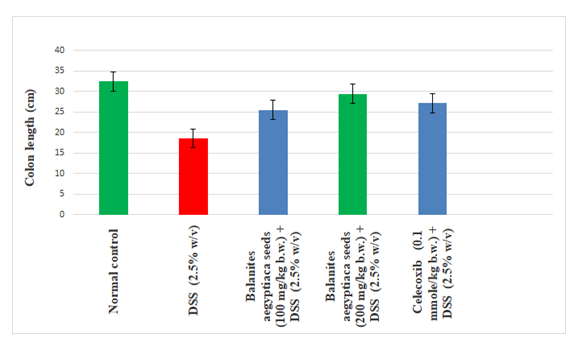
Figure 1:Effect of Balanites aegyptiaca seeds extract and celecoxib on colon length in DSS induced colitis groups of rats. Data was expressed as mean ± SEM (n=6).
Table 2 and 3 show a significantly (P < 0.05) decreased colon levels of GSH, GST while significantly increasing colon TBARS, TNF- α, IFN-γ, IL-1 and IL-6 in normal in the DSS-treated rats as compared with the normal control group (P<0.05), indicating acute colon damage. Balanites aegyptiaca seeds extract (100 and 200 mg/kg.b.w) and celecoxib (0.1mmole/kg.b.w)significantly (P < 0.05) enhanced the colon enzymes activities GST as well as level of GSHin rats and decrease TBARS, TNF- α, IL-1 and IL-6 levels, as compared to the DSS-treated group.
| Group | GSH mg/g protein | GST | TBARS (nmol/mg protein) |
|---|---|---|---|
Normal control A |
9.54 ± 0.78c |
195.98 ± 13.28e |
32.98 ± 5.74a |
DSS (2.5% (w/v) in drinking water positive control |
3.88 ± 0.33a |
110.65 ± 12.06a |
96.53 ± 8.33e |
Balanites aegyptiaca (100 mg/kg b.w.)+ DSS (2.5% (w/v) |
5.80 ± 0.68b |
154.09 ± 8.70b |
52.09 ± 7.80d |
Balanites aegyptiaca (200 mg/kg b.w.)+ DSS (2.5% (w/v) |
9.06 ± 0.95c |
183.25 ± 15.40c |
42.10 ± 3.95b |
Celecoxib (0.1 mmole/kg.b.w.) + DSS (2.5% (w/v) |
8.77 ± 0.55c |
170.65 ± 9.87d |
49.80 ± 5.90c |
Table 2:Levels of colon reduced glutathione (GSH), thiobarbaturic acid reactive substances (TBARS), glutathione-S-transferase (GST) in normal and experimental groups of rats. It was given to all groups except the normal one. The extracts (100, 200 mg/kg.b.w.) were orally given daily for 2 weeks. Values are given as mean ± SD for groups of six animals each. Data followed by the same letter are not significantly different at P ≤ 0.05.
Figure 2 and 3 displayed that DSS (2.5%) promoted the COX-2 and P53 protein expression in DSS- treated the group of rats compared with control group. Administration of Balanites aegyptiaca seeds extract (100 and 200 mg/kg.b.w) and celecoxib (0.1mmole/kg.b.w)significantly (P < 0.05), led to a statistically significant decrease of COX-2 and P53 protein expression relative to DSS treated group of rats after 14 days (p< 0.05). Agarose gel electrophoresis images of COX-2, P53 and β-actin support the present results (Figure 4).
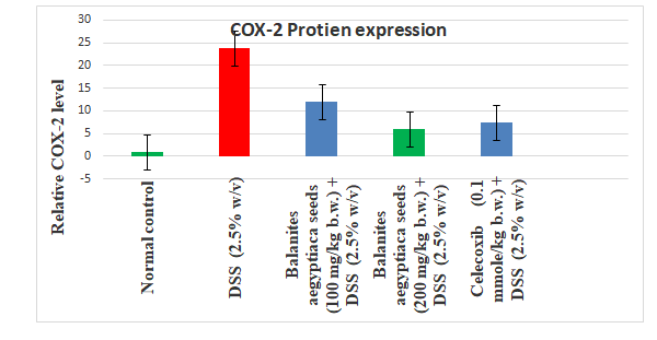
Figure 2:Effect of Balanites aegyptiaca seeds extract and celecoxib on colon COX-2 protein expression in rats. Data (n = 3 per group) are presented as mean ± SD. *p<0.05.
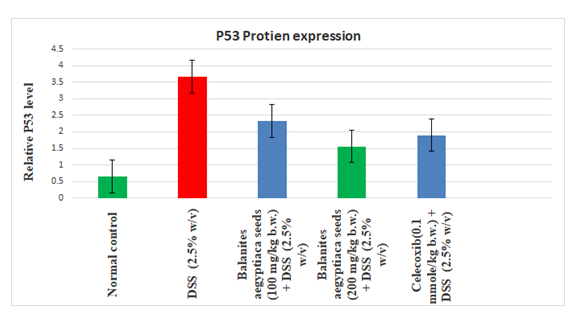
Figure 3:Effect of Balanites aegyptiaca seeds extract and celecoxib on colon P53 protein expression in rats. Data (n= 3 per group) are presented as mean ± SD. *p<0.05.
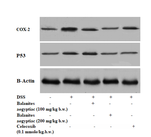
Figure 4:Electrophoretic pattern of Balanites aegyptiaca seeds extract and celecoxib on colon protein expression of COX-2, P53 and B-actin in rats.
Microscopic pictures of H&E stained colon sections showing wide surface ulceration (the black arrow), the glands are irregular with decreased mucin production (blue arrows). The lamina propria showed dense inflammatory cell exudate (the star). The changes are similar to those of ulcerative colitis (Figure 5A) in group received DSS (5%). In contrast, showed intact surface mucosa with regular glands showed adequate mucin secretion, free of remarkable pathological changes (Figure 5C and D) in group received DSS (5%) + aegyptiaca seeds extract (100 and 200 mg/kg.b.w) and/or celecoxib (0.1mmole/kg.b.w).
Ulcerative colitis (UC) is associated with an immune mediated disorder of the intestine and increased cell proliferation and evasion from apoptosis due to various environmental conditions [32]. Also, its associated with overproduction of inflammatory mediators, such as tumor necrosis factor (TNF-alpha) and NF-kB [22,33] which led to increase inflammatory reaction generates excess ROS, activation of proteolytic enzymes and cytokines which lead to tissue damage [22,34].
Induction of UC in animal by oral administration of DSS gave the same histological and immunological changes in human IBD [35].Conventional drugs used for treatment of IBD are mostly anti-inflammatory agents, including celecoxib [25]. Balanites aegyptiaca, a botanical cousin to ginger, has been traditionally used in medicine for treating inflammatory and gastrointestinal disorders [10,15].
In our study, exposure to DSS in drinking water for 14 days, induced acute colitis and the chronic colitis with significantly decreased of colon length in DSS treated group of rats. Mahmoud.,et al. [22] have reported a significant correlation between the DAI score and pathological changes in DSS induced acute and chronic colitis. DSS induced colitic animals exhibited a significant weight loss, developed bloody diarrhea [22].
Balanites aegyptiaca, significantly attenuated this DSS induced UC, and colonic shortening and prevented the anemia due to rectal bleeding. Reduction in the DAI score indicates that Balanites aegyptiaca extract prevents the initial progression of UC.
During DSS induced colitis, the antioxidant status of the colonic tissue was altered and resulted in the increased production of reactive oxygen species (ROS). According to a previous study on the antioxidant enzyme profile in a DSS induced model, a marked depletion of the cellular antioxidant GSH was observed [22]. Clinical studies on IBD also revealed the imbalance in antioxidant status [36]. The antioxidant profile was restored by Balanites aegyptiaca extract treatment, which suggested its anti-oxidant property. Also, the antioxidant and anti-inflammatory activity of Balanites aegyptiaca was proved in previous studies [10-15].
Under homeostatic conditions, the intestinal mucosa can maintain the balance between proinflammatory and anti-inflammatory cytokines. However, IBD patients often display increased intestinal permeability and impaired epithelial barrier function [37], leading to elevated levels of pro-inflammatory cytokines such TNF-α, IFN-γ, interleukin (IL)-1, IL-6, and IL-12 [38]. Similar increases in pro-inflammatory mediators are also observed in the DSS-colitis model correlating well with clinical parameters [38]. Consistent with the known anti-inflammatory effects of some probiotic strains [39,40], we found that Balanites aegyptiacasignificantly reduced levels of colon tumor necrosis factor-α (TNF-α), interferon-γ (IFN-γ), interleukin (IL)-1 and interleukin (IL)-6 in the colons of treated mice, which were accompanied by increased anti-inflammatory cytokine expression COX-2and P53.
NF-kB has a specific role in coupling inflammation to cancer by regulating the functions of inflammatory gene expression such as TNF-alpha and IL-6. DSS has a direct toxic effect on the epithelium and destroys the mucosal barrier and causes the elevated secretion of pro-inflammatory cytokines such as TNF-alpha [22].
Increased expression of P53 and COX-2 has been reported in many colorectal tumors and adenocarcinomas. COX-2 expression increases from normal to progression of cancerous state.Our result clearly indicates that the Balanites aegyptiaca markedly inhibited P53 and COX-2 mRNA expressions in DSS treated rats. Histological examination of colonic sections revealed an altered architecture of colon mucosa with typical inflammatory changes in colonic architecture such as crypt and surface epithelial loss as well as infiltration of inflammatory cells and complete destruction of the epithelial architecture. The severity of the disease increases with chronicity. These macroscopic and microscopic alterations observed in our study agreed with numerous studies reported on DSS induced UC models in rats [22]. Balanites aegyptiaca led to marked reduction in the inflammatory infiltrate in both lamina propria and submucosa and dose-dependently protected against changes in colon length and mass index.
In the present study, the histological findings prove that resveratrol affected the recovery of the colon structure in rats with DSS -induced colon toxicity. Indeed, there was free of remarkable pathological changes in rats treated with Balanites aegyptiacagroups compared to the control DSS treated group. Histological studies confirmed the colon protective effect of Balanites aegyptiaca.
Present data evidence that secreted colon tumor necrosis factor-α (TNF-α), interferon-γ (IFN-γ), interleukin (IL)-1 and interleukin (IL)-6 is a proinflammatory cytokine and an active protagonist of gut mucosal inflammation. In this study, we explored in vivo the potential of Balanites aegyptiaca, a P53 and COX-2 inhibitor, as an anti-inflammatory medicinal plant that showed a strongly ameliorates DSS-induced colitis in rats. We reason that Balanites aegyptiaca may represent an innovative tool for the management of human intestinal inflammation.
Citation: Mohammed A Hussein., et al. “Balanites aegyptiaca Extract Inhibits COX-2 and P53 Expression in DSS-induced Ulcerative Colitis". Acta Scientific Nutritional Health 5.5 (2021): 120-127.
Copyright: © 2021 Mohammed A Hussein., et al. This is an open-access article distributed under the terms of the Creative Commons Attribution License, which permits unrestricted use, distribution, and reproduction in any medium, provided the original author and source are credited.


 Open Access by
Acta Scientific is licensed under a Creative Commons Attribution 4.0 International License
Open Access by
Acta Scientific is licensed under a Creative Commons Attribution 4.0 International License
Based on a work at https://actascientific.com
ff
