Hazar Khankan* and Sawssan Ali
Pulmonology Division, Department of Pediatrics, Children's Damascus University Hospital, Syria
*Corresponding Author: Hazar Khankan, Pulmonology Division, Department of Pediatrics, Children's Damascus University Hospital, Syria.
Received: April 29, 2019; Published: July 10, 2019
Citation: Hazar Khankan and Sawssan Ali. “Congenital Pulmonary Airway Malformation Type I: A Rare Case of an Infant”. Acta Scientific Paediatrics 2.8 (2019):28-29.
A 1-month-old female infant presented to Children's Damascus University Hospital with a history of tachypnea for about 3 weeks.
On physical examination, she had mild intercostal retractions and crackles on both sides of the chest with slightly diminished breath sounds on the right side. Respiratory rate was about 40 and oxygen saturation was 86% on room air.
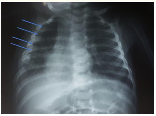
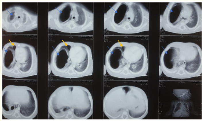
On investigations, chest X-ray showed large hyperlucent area in the right lung with poor lung markings. CECT images showed a large cystic lesion occupying most of the right upper lobe with a smaller cyst in the right middle lobe, a compression in the right lower lobe with a small amount of pleural effusion in the same side, and a mediastinal shift to the left.
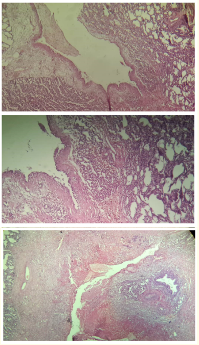
Lobectomy was performed and the resected specimen sent to histopathological study. It revealed small immature rudimentary alveoli, some are dilated, with presence of multiloculated cysts up to 3 cm, lined by ulcerated columnar to pseudostratified epithelium and surrounded by severe chronic inflammation, interstitial hemorrhage, granulation tissue formation and fibrosis. Features were compatible with congenital pulmonary airway malformation type 1.

Image 1: CXR: Hyperlucent area in the right lung (Blue arrows).

Image 2: CECT: Large right upper cyst (Blue arrows). Small right middle cyst (yellow arrows).

Image 3: The microscopic appearance.
Special thanks to the Pathologist Dr. Lina Al Haffar, MD.
Copyright: © 2019 : Hazar Khankan and Sawssan Ali. This is an open-access article distributed under the terms of the Creative Commons Attribution License, which permits unrestricted use, distribution, and reproduction in any medium, provided the original author and source are credited.
ff
© 2024 Acta Scientific, All rights reserved.