Yousef Shanti1, Ibrahim Taha2*, Adnan Busttami3, Hamzeh Al Zabadi4, Saif-eddin Malhas3, Qutaiba Mahmoud3, Rami Othman5, Liana Al-Labadi6 and Alshaarawi Salem7
1Ophthalmology Department, An-Najah National University Hospital, Nablus, Palestine
2Department of Translational Medicine, University of Pavia, Italy
3Faculty of Medicine and Health Sciences, An-Najah National University, Nablus,
Palestine
4Public Health Department, An-Najah National University, Nablus, Palestine
5Bachelor of Medical Laboratory Science, An-Najah National University, Nablus,
Palestine
6Optometry Department, Arab American University, Palestine
7Ph.D. Optometry and vision sciences, University of Minho, Portugal
*Corresponding Author: Ibrahim Taha, Department of Translational Medicine, University of Pavia, Italy.
Received: February 01, 2021; Published: February 19, 2021
Background: Cataract is a major cause of visual impairment among diabetic patients. The study aimed to assess the prevalence of cataracts and its associated factors among diabetes mellitus patients in the West Bank, Palestine, in order to better identify the targets for future healthcare policy and interventions.
Materials and Methods: A prospective quantitative cross-sectional study was conducted in all governorates of the West Bank with a proportionally and randomly selected sample size of 385 subjects. The participants were interviewed face to face for an average of ten minutes to fill in a previously validated Early Treatment Diabetic Retinopathy Study (ETDRS) questionnaire. Then after, blood and urine samples were collected and finally, the participants were passed for an ophthalmic examination performed by a certified ophthalmologist.
Results: The prevalence of any cataract, nuclear cataract, cortical cataract, and posterior subcapsular cataract was found to be 47.8%, 28.1%, 14.0%, and 4.7% respectively.
The most significant factors for any cataract were: age (55 - 64) (OR 5.375), age (≥ 65) (OR 30.238), DM duration (10 - 19) (OR 2.251), DM duration (≥ 20) (OR 5.771), systemic steroid therapy (OR 0.411). For nuclear cataract, significant associated factors were age (55 - 64) (OR 9.576), age (≥ 65) (OR 17.928), DM duration (≥ 20) (OR 3.059), ALK PHOS (OR 0.354).
For cortical cataract: DM duration (≥ 20) (OR 6.134) was the only significant factor. And in the case of posterior subcapsular cataract, age was the only significant factor (OR 1.076).
Conclusion: The high prevalence of cataract among diabetic patients in West Bank indicates the importance of cataract screening program for DM patients as an attempt to prevent visual impairment and related disabilities.
Keywords: Diabetes Mellitus; Cataract; Blindness
NC: Nuclear Cataract; CC: Cortical Cataract; PSC: Posterior Subcapsular Cataract; DM: Diabetes Mellitus; MoH: Ministry of Health; PHC: Primary Health Care; BMI: Body Mass Index; HbA1c: Glycated Hemoglobin; TG: Triglycerides; CHOL: Cholesterol; HDL: High Density Lipoprotein; LDL: Low Density Lipoprotein; ALK PHOS: Alkaline Phosphatase; GOT: Glutamic-Oxaloacetic Transaminase; GPT: Glutamic-Pyruvic Transaminase; CREAT: Creatinine; BU: Blood; MLAB: Microalbuminuria; SD: Standard Deviation; OR: Odds Ratio, CI: Confidence Interval
Cataract is the most common cause of visual impairment worldwide. It has a significant contribution to blindness mainly in developing countries [1]. Cataract is responsible for about 51% of worldwide blindness [2].
Cataract is a common ophthalmologic condition in which the normally clear lens of the eye becomes progressively opaque. As the function of the lens is to pass and focus the light onto the retina, any significant opacity in the lens would deviate and scatter the light and prevent a sharply defined image from reaching the retina resulting in visual impairment [3]. The pathogenesis of cataract is multi-factorial and not fully understood, but there are many hypotheses to explain the pathogenesis which include: compaction and stiffening of the lens material, abnormal changes in lens proteins, pigmentation of lens proteins and changes in the ionic components of the lens [4].
Risk factors of cataract are usually related to the general health of the eyes in addition to uncontrolled Diabetes Mellitus (DM), steroid therapy (oral, intravenous, inhaled or topical), smoking, ocular trauma, previous ocular surgery and genetic predisposition [5].
Cataract can be classified into acquired and congenital. Acquired sub classification include age related cataract (the most common type of cataract), systemic diseases related cataract; diabetes mellitus, traumatic cataract; physical, chemical and radiation. According to the anatomy of human lens and cataract location, there are 3 common types: Nuclear Cataract (NC), Cortical Cataract (CC), and Posterior Subcapsular Cataract (PSC) [5].
DM is one of the most common chronic non-communicable diseases worldwide, the prevalence is increasing due to population growth, aging, urbanization and increasing prevalence of obesity and physical inactivity [6]. Globally, in 2015 more than 415 million people were affected by DM and the number is estimated to reach around 642 million by 2040 according to the International Diabetes Federation [7]. In the Eastern Mediterranean Region, there were 15,188,000 diabetic cases in the year 2000 and it is estimated to reach around 42,600,000 cases in 2030 [8]. In West Bank in Palestine, there were 278,302 patients in 2019 with a total population of 2,862,485 [9].
As the incidence and progression of cataract is increased in patients with diabetes mellitus, cataract is a major cause of visual impairment among diabetic patients [10,11]. The pathogenesis of cataract among DM patients is not fully understood but many hypotheses support that the mechanism of cataract formation due to generation of polyols from glucose by aldose reductase enzyme, which result in increased osmotic pressure in the lens fibers and subsequent swelling and rupture of the lens [12]. DM patients have increased risk of cataract formation compared to non DM population [13,14]. Age, longer duration of DM, increased level of glycated hemoglobin (HbA1c), severity of diabetic retinopathy, proteinuria and DM treatment were all considered as associated factors [13]. CC and PSC cataract were found to be associated with DM while NC was associated with elevated HbA1c level [15].
The surgical treatment is usually indicated before the maturity of the cataract to a level that will prevent the full fundus examination [13]. Cataract surgery in DM compared to non-DM patients have poorer vision outcome and more complications such as acceleration of retinopathy, rubeosis and macular changes like macular edema or cystoid macular edema [16,17].
This study aimed to assess the prevalence of cataract and identify its mostly associated factors among the Palestinian DM patients in the West Bank directorates in order to target the effective screening programs that would minimize the progression of cataract or delay its formation.
A prospective quantitative cross-sectional study was conducted. The study population of DM patients in West Bank in 2019 was estimated to be around 278,302 patients according to the MoH [9]. Subjects were selected from the DM patients who were above 18 years of age and were followed up in Palestinian ministry of health (MoH) centers. Subjects were recruited in the primary health care centers (PHC) of the MoH in all West Bank directorates (Jenin, Tulkarm, Qalqelia, Nablus, Salfit, Tubas, Ramallah, Bethlehem, Jericho, and Hebron). PHC centers of MoH were chosen due to their accessibility and representativeness to the majority of DM patients in the West Bank. In addition, 422 of 608 PHC centers are provided by MoH so it is considered as the major healthcare provider for DM patients in the West Bank and the vast majority of DM patients seek healthcare in the governmental sector as those patients are usually insured, it should be noted that 189916 families are having governmental insurance [9]. DM patients with cognitive dysfunction or refused to participate were not included.
Sample sizeSubjects were recruited by a random proportional method. The sample size was calculated using the following equation [18]:
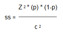
Z = Standard normal variate (1.96 for 95% confidence level) p = Expected proportion in population. c = Absolute error.
We used a standard normal variate of 1.96 and an absolute error of 5%. However, there were no previous research in Palestine regarding this topic so we assume the expected proportion in population (p) to be 0.5 in order to have the highest sample size. After inserting these figures into the previously mentioned equation, the total sample was 385. Distribution of sample between West Bank directorates depended on proportion of each directorate DM patients from the total West Bank DM patients excluding those under 18 years old [9]. Therefore, 83 patients were chosen from Nablus, 77 patients from Hebron, 58 patients from Jenin, 48 patients from Tulkarm, 39 patients from Ramallah, 24 patients from Bethlehem, 17 patients from Qalqelia, 16 patients from Jerusalem, 9 patients from Salfit, 9 patients from Tubas, 5 patients from Jericho [8].
DefinitionsSubjects were first recruited. The procedure of recruitment was as following: a list of diabetic patients in each directorate was obtained from MoH, subjects were randomly selected from each directorate by using random sampling technique. After that, subjects were called by phone to tell them about participation in this research, subjects were motivated to participate by telling them that their participation will be free screening for cataract and lab tests will be done by qualified team. Subjects in each directorate were told about the time and PHC center which is the main center for directorate.
The selected subjects were interviewed face to face for an average of ten minutes to fill the study’s previously modified validated Early Treatment Diabetic Retinopathy Study (ETDRS) questionnaire. Then, blood and urine sample were obtained and then after, subjects underwent pupil dilation by using a mydriatic agent (Mydramide is a Tropicamide 5% which produced by Dr. Fischer company), one drop for each eye. Twenty to thirty minutes after that, dilated eye exam done by the ophthalmologist using Slit-Lamp and then the results were documented based on the Lens Opacities Classification System II (LOCS II) [19].
The modified questionnaire was composed of 5 sections; (1) socio-demographic data including age, sex and directorate; (2) medical history including weight and height, DM type, DM duration and treatment, history of hypertension, history of systemic steroid therapy; (3) ocular history including ocular trauma or topical steroid therapy or retinopathy treatment; (4) ophthalmic examination results using slit-lamp according to ophthalmologist findings regarding cataract and its types and grades if present; (5) blood, serum and urine laboratory tests performed by the research team in Nablus city Charitable Medical Complex. Laboratory tests included: Glycated Hemoglobin (HbA1c), Triglycerides (TG), Cholesterol (CHOL), High Density Lipoprotein (HDL), Low Density Lipoprotein (LDL), Alkaline Phosphatase (ALK PHOS), Glutamic-Oxaloacetic Transaminase (GOT), Glutamic-Pyruvic Transaminase (GPT), Creatinine (CREAT), Blood Urea (BU), and Microalbumuria (MALB).
Laboratory proceduresBlood and urine samples were obtained from patients according to the recommendations of manufacturer of used kits. Blood sample was taken at fasting status for 8 - 10 hours by vacutainer blood tubes (4 ml clot activator tube and 3 ml EDTA tube) for each patient, urine sample was taken as random midstream around 50 ml volume. All samples were stored by sample transporter box at temperature 2 - 8 Celsius till reached the lab, where urine and clot activator samples were centrifuged to prepare urine and serum for tests.
EDTA tube whole blood was analyzed by HumaMeter A1c system which is a product of Human Germany, Boronate Affinity Quenching Technology is based on affinity of boronate for glycated proteins, fluorescent binding and quenching which have measuring range: 4 - 15% and imprecision: < 3%.
Serum and urine samples were analyzed by HumaStar 200 system which is a product of Human Germany, which is an open fully automated chemistry analyzer awarded the international iF product design award.
Method of each test used as the following:
All kits used are produced by Human Germany according to the recommendations of the used systems. The normal cutoff values for each test were obtained from kits sheets as the following: CHOL ≤ 190 mg/dl. TG ≤ 150. HDL ≥ 60 mg/dl. LDL ≤ 129 mg/dl. ALK PHOS ≤ 104 U/L, 129 U/L for female, male respectively. GOT ≤ 31 U/L, 35 U/L for female, male respectively. GPT ≤ 34 U/L, 45 U/L for female, male respectively. BU ≤ 50 mg/dl. CREAT ≤ 0.9 mg/dl, 1.1 mg/dl for female, male respectively. MLAB 0-30 mg/l. HbA1c ≤ 7% (NGSP5/DCCT6) for glycemic control.
Internal and external controls were used at the start and at the end of each run of samples to maximize the accuracy of results.
Internal control by using recommended Human controls kits (Serodos and Serodos Plus for serum tests, Mircoalbumin Standard for urine test and HumaMeter A1c control for HbA1c). External control by comparison of HbA1c results with HPLC method (D10) from BioRad.
Data analysisStatistical Product and Service Solutions (SPSS V.22, IBM SPSS statistic) was used for data entry and analysis.
Descriptive statistical analysis was used to determine the Mean ± SD for numerical variables, in addition to the frequency and percentage for categorical variables.
Univariate analysis using Chi-square testing was used to examine the association between various factors (sociodemographic, medical history, ocular history, laboratory tests) and the dependent variables (AC, NC, CC, PSC), in which the association was considered to be statistically significant at P ˂ 0.05.
Multivariate analysis using Binary logistic regression was used to determine the adjusted association between independent variables (that was found to be significant using the univariate analysis) and dependent variables, to address the confounding and produce adjusted OR.
The total number of participants in the study was 385, with a response rate 100%.
PrevalenceThe prevalence of AC among diabetic patients was found to be 47.8%. While the prevalence of NC was 28.1%, CC 14.0% and PSC 4.7% (Table 1).
| Variable | Frequency (%) |
|---|---|
AC1 Absent Present |
201 (52.2) 184 (47.8) |
NC2 Absent Present |
277 (71.9) 108 (28.1) |
CC3 Absent Present |
331 (86.0) 54 (14.0) |
PSC4 Absent Present |
367 (95.3) 18 (4.7) |
Table 1: Prevalence of AC, NC, CC and PSC (N = 385).
1: Any Cataract, 2: Nuclear Cataract, 3: Cortical Cataract, 4: Posterior Subcapsular Cataract.
Sociodemographic factors: The mean age of participants was 56.48 ± 12.337 years, the predominant age category was 55 - 64 with 36.4% of participants. 52.7% of them were females. 56.1% of participants were from North West Bank. More than half of participants, 51.9% had had primary education (Table 2).
| Variable | Frequency (%) |
|---|---|
Age category ≤ 44 45 - 54 55 - 64 ≥ 65 |
45 (11.7) 100 (26.0) 140 (36.4) 100 (26.0) |
Sex Female Male |
203 (52.7) 182 (47.3) |
Directorate category North West Bank Middle West Bank South West Bank |
216 (56.1%) 68 (17.7) 101 (26.2) |
Education level Not educated Primary education Secondary education High education |
37 (9.6) 200 (51.9) 79 (20.5 69 (17.9) |
Age (Mean ± SD) |
56.48 ± 12.337 |
Table 2: Socio-demographic characteristics of participants (N = 385).
Medical history: The BMI mean was 30.3349 ± 5.59523, 38.4% of them were over weighted whereas 49.6% were obese. DM duration mean was 9.69 ± 7.866 years, Type 2 DM was predominant among participants with 92.5% whereas 7.5% of participants had Type 1 DM. 46.2% of participants were treated with oral hypoglycemic agents, 25.5% with insulin and 17.4% with a combination of both oral hypoglycemic agents and insulin. 55.3% had hypertension, 12.7% had a history of systemic steroid therapy. Only 20.5% were current smokers (Table 3).
| Variable | Frequency (%) |
|---|---|
BMI1 category Under weight Normal weight Overweight Obesity |
2 (0.5) 44 (11.4) 148 (38.4) 191 (49.6) |
DM2 type Type 1 DM Type 2 DM |
29 (7.5) 356 (92.5) |
DM2 duration category ≤ 4 5 - 9 10 - 19 ≥ 20 |
126 (32.7) 78 (20.3) 132 (34.3) 49 (12.7) |
DM2 treatment No treatment Diet Oral hypoglycemic agents Insulin Diet and oral hypoglycemic agents Diet and insulin Oral hypoglycemic agents and insulin Diet and oral hypoglycemic agents and insulin |
3 (0.8) 12 (3.1) 178 (46.2) 98 (25.5) 16 (4.2) 6 (1.6) 76 (17.4) 5 (1.3) |
Hypertension Absent Present |
172 (44.7) 213 (55.3) |
Current smoking No Yes |
306 (79.5) 79 (20.5) |
Systemic steroid therapy No Yes |
336 (87.3) 49 (12.7) |
BMI1 (Mean ± SD) |
30.3349 ± 5.59523 |
DM2 duration (Mean ± SD) |
9.69 ± 7.866 |
Table 3: Medical characteristics of participants (N = 385).
1: Diabetes Mellitus, 2: Body Mass Index.
Ocular history: 10.6% of participants had previous ocular trauma. 6% were previously treated with ocular topical steroid and 14.8% had treatment for retinopathy using various medical and surgical methods (Table 4).
| Variable | Frequency (%) |
|---|---|
Ocular trauma No Yes |
344 (89.4) 41 (10.6) |
Topical steroid therapy No Yes |
362 (94.0) 23 (6.0) |
Retinopathy treatment No Injection LASER Surgery Injection and LASER Injection and surgery LASER and surgery Injection and LASER and surgery |
328 (85.2) 5 (1.3) 17 (4.4) 17 (4.4) 8 (2.1) 2 (0.5) 3 (0.8) 5 (1.3) |
Table 4: Ocular characteristics of participants (N = 385).
Laboratory tests: The mean of HbA1c was 8.2229 ± 1.86130%, 68.1% of participants had non-controlled HbA1c level. The mean of TG was 189.5844 ± 132.33346 mg/dl, 50.1% of them had abnormal TG level. The mean of HDL was 48.6512 ± 14.51382 mg/dl, 83.9% of them had abnormal level. The mean of ALK PHOS was 229.8571 ± 114.79696 U, 93.8% of them had abnormal level. The mean of CREAT was 1.0285 ± 0.47192 mg/dl, 41.0% of them had abnormal level. The mean of BU was 34.2735 ± 18.88311 mg/dl, 12.7% of them had abnormal level. The mean of MLAB was 68.5577 ± 78.17979 mg/dl, with 54.8% had microalbuminuria and only 2.1% had macroalbuminuria (Table 5).
| Variable | Frequency (%) |
|---|---|
HbA1c1 level Controlled Non - controlled |
123 (31.9) 262 (68.1) |
TG2 level Normal Abnormal |
192 (49.9) 193 (50.1) |
CHOL3 level Normal Abnormal |
236 (61.3) 149 (38.7) |
HDL4 level Normal Abnormal |
62 (16.1) 323 (83.9) |
LDL5 level Normal Abnormal |
282 (73.2) 103 (26.8) |
ALK PHOS6 level Normal Abnormal |
24 (6.2) 361 (93.8) |
GOT7 level Normal Abnormal |
313 (81.3) 72 (18.7) |
GPT8 level Normal Abnormal |
358 (93.0) 27 (7.0) |
CREAT9 level Normal Abnormal |
227 (59.0) 158 (41.0) |
BU10 level: Normal Abnormal |
336 (87.3) 49 (12.7) |
Albuminuria11 Normal Microalbuminuria Microalbuminuria Macroalbuminuria |
166 (43.1) 211 (54.8) 8 (2.1) |
HbA1c1 (Mean ± SD) |
8.2229 ± 1.86130 |
TG2 (Mean ± SD) |
189.5844 ± 132.33346 |
CHOL3 (Mean ± SD) |
181.8442 ± 47.46777 |
HDL4 (Mean ± SD) |
48.6512 ± 14.51382 |
LDL5 (Mean ± SD) |
106.1665 ± 39.42505 |
ALK PHOS6 (Mean ± SD) |
229.8571 ± 114.79696 |
GOT7 (Mean ± SD) |
26.3143 ± 10.91169 |
GPT8 (Mean ± SD) |
23.4468 ± 9.88280 |
CREAT9 (Mean ± SD) |
1.0285 ± 0.47192 |
BU10 (Mean ± SD) |
34.2735 ± 18.88311 |
Albuminuria11 (Mean ± SD) |
68.5577 ± 78.17979 |
Table 5: : Laboratory tests characteristics of participants (N = 385).
1: Glycated Hemoglobin, 2: Triglycerides, 3: Cholesterol, 4: High Density Lipoprotein, 5: Low Density Lipoprotein, 6: Alkaline Phosphatase, 7: Glutamic-Oxaloacetic Transaminase, 8: Glutamic-Pyruvic Transaminase, 9: Creatinine, 10: Blood Urea, 11: Albuminuria.
Sociodemographic factors: The results showed a statistically significant association between age and AC (P ˂ .001), age and NC (P ˂ .001), age and CC (P = .015), age and PSC (P = .044) (Table 6).
| Variable | AC1 | NC2 | CC3 | PSC4 | ||||||||
|---|---|---|---|---|---|---|---|---|---|---|---|---|
No % |
Yes % |
P value |
No % |
Yes % |
P value |
No % |
Yes % |
P value |
No % |
Yes % |
P value |
|
Age category ≤ 44 45 - 54 55 - 64 ≥ 65 |
88.9 72.0 51.4 17.0 |
11.1 28.0 48.6 83.0 |
˂ .001 |
97.8 84.0 68.6 53.0 |
2.2 16.0 31.4 47.0 |
˂ .001 |
91.1 93.0 85.0 78.0 |
8.9 7.0 15.0 22.0 |
.015 |
100.0 98.0 95.0 91.0 |
0.0 2.0 5.0 9.0 |
.044 |
Sex Female Male |
54.7 49.5 |
45.3 50.5 |
.305 |
74.4 69.2 |
25.6 30.8 |
.261 |
86.7 85.2 |
13.3 14.8 |
0.665 |
94.6 96.2 |
5.4 3.8 |
.466 |
Directorate category North West Bank Middle West Bank South West Bank |
49.1 52.9 58.4 |
50.9 47.1 41.6 |
.297 |
69.4 73.5 76.2 |
30.6 26.5 23.8 |
.433 |
85.6 83.8 88.1 |
14.4 16.2 11.9 |
.717 |
94.9 97.1 95.0 |
5.1 2.9 5.0 |
.865 |
Table 6: : Univariable analysis of sociodemographic characteristics and cataract.
1: Any Cataract, 2: Nuclear Cataract, 3: Cortical Cataract, 4: Posterior Subcapsular Cataract.
Medical history: The results showed a statistically significant association between DM type and AC (P =.002), DM type and NC (P =.002), DM duration and AC (P ˂ .001), DM duration and NC (P =.003), DM duration and CC (P ˂ .001), hypertension and AC (P ˂ .001), hypertension and NC (P =.001), hypertension and PSC (P =.014), systemic steroid therapy and AC (P =.023) (Table 7).
| Variable | AC1 | NC2 | CC3 | PSC4 | ||||||||
|---|---|---|---|---|---|---|---|---|---|---|---|---|
No % |
Yes % |
P value |
No % |
Yes % |
P value |
No % |
Yes % |
P value |
No % |
Yes % |
P value |
|
BMI5 group Underweight Normal weight Overweight Obesity |
0.0 59.1 48.0 54.5 |
100.0 40.9 52.0 45.5 |
.249 |
0.0 72.7 70.9 73.3 |
100.0 27.3 29.1 26.7 |
.213 |
50.0 81.8 82.4 90.1 |
50.0 18.2 17.6 9.9 |
.060 |
100.0 93.2 95.9 95.3 |
0.0 6.8 4.1 4.7 |
.753 |
DM6 type Type 1 DM Type 2 DM |
79.3 50.0 |
20.7 50.0 |
.002 |
96.6 69.9 |
3.4 30.1 |
.002 |
79.3 86.5 |
20.7 13.5 |
.271 |
96.6 95.2 |
3.4 4.8 |
1.000 |
DM5 duration category ≤ 4 5 - 9 10 - 19 ≥ 20 |
71.4 56.4 43.2 20.4 |
28.6 43.6 56.8 79.6 |
˂ .001 |
81.7 75.6 68.2 51.0 |
18.3 24.4 31.8 49.0 |
˂ .001 |
93.7 94.9 81.8 63.3 |
6.3 5.1 18.2 36.7 |
˂ .001 |
96.8 97.4 93.9 91.8 |
3.2 2.6 6.1 8.2 |
.335 |
Hypertension Absent Present |
62.8 43.7 |
37.2 56.3 |
˂ .001 |
80.2 65.3 |
19.8 34.7 |
.001 |
89.5 83.1 |
10.5 16.9 |
.071 |
98.3 93.0 |
1.7 7.0 |
.014 |
Current smoking No Yes |
52.0 53.2 |
48.0 46.8 |
.849 |
72.5 69.6 |
27.5 30.4 |
.605 |
85.9 86.1 |
14.1 13.9 |
.977 |
94.4 98.7 |
5.6 1.3 |
.139 |
Systemic steroid therapy No Yes |
50.0 67.3 |
50.0 32.7 |
.023 |
71.1 77.6 |
28.9 22.4 |
.350 |
85.1 91.8 |
14.9 8.2 |
.206 |
94.6 100.0 |
5.4 0.0 |
.145 |
Table 7: : Univariable analysis of medical characteristics and cataract.
1: Any Cataract, 2: Nuclear Cataract, 3: Cortical Cataract, 4: Posterior Subcapsular Cataract, 5: Body Mass Index, 6: Diabetes Mellitus.
Ocular history: The results showed a statistically significant association between ocular trauma and NC (P = 0.043), retinopathy treatment and AC (P ˂ .001), retinopathy treatment and NC (P ˂ .001), retinopathy treatment and CC (P = 0.003) (Table 8).
| Variable | AC1 | NC2 | CC3 | PSC4 | |||||||||
|---|---|---|---|---|---|---|---|---|---|---|---|---|---|
No % |
Yes % |
P value |
No % |
Yes % |
P value |
No % |
Yes % |
P value |
No % |
Yes % |
P value |
||
Ocular trauma No Yes |
52.0 53.7 |
48.0 46.3 |
.844 |
70.3 85.4 |
29.7 14.6 |
.043 |
85.5 90.2 |
14.5 9.8 |
.405 |
95.3 95.1 |
4.7 4.9 |
1.000 |
|
Topical steroid therapy No Yes |
53.0 39.1 |
47.0 60.9 |
.195 |
71.5 78.3 |
28.5 21.7 |
.487 |
86.2 82.6 |
13.8 17.4 |
.546 |
95.3 95.7 |
4.7 4.3 |
1.000 |
|
Retinopathy treatment No Injection LASER Surgery Injection and LASER Injection and surgery LASER and surgery Injection and LASER and surgery |
57.6 0.0 29.4 17.6 37.5 0.0 0.0 20.0 |
42.4 100.0 70.6 82.4 62.5 100.0 100.0 80.0 |
˂ .001 |
73.2 20.0 41.2 100.0 37.5 100.0 66.7 100.0 |
26.8 80.0 58.8 0.0 62.5 0.0 33.3 0.0 |
˂ .001 |
88.1 40.0 70.6 94.1 62.5 50.0 66.7 80.0 |
11.9 60.0 29.4 5.9 37.5 50.0 33.3 20.0 |
.003 |
96.0 60.0 88.2 94.1 100.0 100.0 100.0 100.0 |
4.0 40.0 11.8 5.9 0.0 0.0 0.0 0.0 |
.106 |
|
Table 8: Univariable analysis of ocular characteristics and cataract.
1: Any Cataract, 2: Nuclear Cataract, 3: Cortical Cataract, 4: Posterior Subcapsular Cataract.
Laboratory tests: The results showed a statistically significant association between: HbA1c level and CC (P = .049), HDL level and NC (P = .049), ALK PHOS and NC (P = .045), CREAT level and AC (P ˂ .001), CREAT level and NC (P = .014), BU level and AC (P = .020), BU and NC (P = .014) (Table 9).
| Variable | AC1 | NC2 | CC3 | PSC4 | |||||||||
|---|---|---|---|---|---|---|---|---|---|---|---|---|---|
| No % | Yes % | P value | No % | Yes % | P value | No % | Yes % | P value | No % | Yes % | P value | ||
HbA1c5 level Controlled Non - controlled |
57.7 49.6 |
42.3 50.4 |
.138 |
77.2 69.5 |
22.8 30.5 |
0.114 |
91.1 83.6 |
8.9 16.4 |
.049 |
95.9 95.0 |
4.1 5.0 |
.698 |
|
TG6 level Normal Abnormal |
55.7 48.7 |
44.3 51.3 |
.168 |
76.0 67.9 |
24.0 32.1 |
0.075 |
88.0 83.9 |
12.0 16.1 |
.249 |
96.4 94.3 |
3.6 5.7 |
.340 |
|
CHOL7 level Normal Abnormal |
52.1 52.3 |
47.9 47.7 |
.965 |
72.0 71.8 |
28.0 28.2 |
.962 |
87.7 83.2 |
12.3 16.8 |
.230 |
95.3 95.3 |
4.7 4.7 |
.987 |
|
HDL8 level Normal Abnormal |
61.3 50.5 |
38.7 49.5 |
.118 |
82.3 70.0 |
17.7 30.0 |
0.049 |
87.1 85.8 |
12.9 14.2 |
.781 |
98.4 94.7 |
1.6 5.3 |
.328 |
|
LDL9 level Normal Abnormal |
52.5 51.5 |
47.5 48.5 |
.858 |
71.6 72.8 |
28.4 27.2 |
.819 |
86.9 83.5 |
13.1 16.5 |
.397 |
95.4 95.1 |
4.6 4.9 |
1.000 |
|
ALK PHOS10 level Normal Abnormal |
37.5 53.2 |
62.5 46.8 |
.136 |
54.2 73.1 |
45.8 26.9 |
.045 |
91.7 85.6 |
8.3 14.4 |
.553 |
87.5 95.8 |
12.5 4.2 |
.093 |
|
GOT11 level Normal Abnormal |
52.1 52.8 |
47.9 47.2 |
.914 |
71.2 75.0 |
28.8 25.0 |
.523 |
85.9 86.1 |
14.1 13.9 |
.970 |
96.2 91.7 |
3.8 8.3 |
.120 |
|
GPT12 level Normal Abnormal |
51.1 66.7 |
48.9 33.3 |
.119 |
70.9 85.2 |
29.1 14.8 |
.112 |
85.8 88.9 |
14.2 11.1 |
1.000 |
95.5 92.6 |
4.5 7.4 |
.365 |
|
CREAT13 level Normal Abnormal |
59.9 41.1 |
40.1 58.9 |
˂ .001 |
76.7 65.2 |
23.3 34.8 |
.014 |
88.5 82.3 |
11.5 17.7 |
.081 |
96.0 94.3 |
4.0 5.7 |
.429 |
|
BU14 level Normal Abnormal |
54.5 36.7 |
45.5 63.3 |
.020 |
74.1 57.1 |
25.9 42.9 |
.014 |
86.0 85.7 |
14.0 14.3 |
.955 |
95.8 91.8 |
4.2 8.2 |
.265 |
|
Albuminuria15 level Normal Micro range Macro range |
56.0 49.8 37.5 |
44.0 50.2 62.5 |
.348 |
72.9 71.6 62.5 |
27.1 28.4 37.5 |
.802 |
86.7 84.8 100.0 |
13.3 15.2 0.0 |
.446 |
95.2 95.3 100.0 |
4.8 4.7 0.0 |
.818 |
|
Table 9: Univariable analysis of laboratory characteristics and cataract.
1: Any Cataract, 2: Nuclear Cataract, 3: Cortical Cataract, 4: Posterior Subcapsular Cataract, 5: Glycated Hemoglobin, 6: Triglycerides, 7: Cholesterol, 8: High Density Lipoprotein, 9: Low Density Lipoprotein, 10: Alkaline Phosphatase, 11: Glutamic-Oxaloacetic Transaminase, 12: Glutamic-Pyruvic Transaminase, 13: Creatinine, 14: Blood, 15: Albuminuria.
After applying binary logistic regression model on each factor associated with any cataract or cataract types, the result showed.
Age (≥ 65) (P ˂ .001), DM duration (10 - 19) (P = .01), DM (≥ 20) (P ˂ .001), and systemic steroid therapy (P = 0.019) were still to be statistically significant with AC and the rest were not statistically significant. Although Age (45 - 54) (OR 3.276, 0.970 - 11.061) and CREAT level (OR 1.714, 0.989 - 2.970) were associated with increased odds of AC, they were not statistically significant (Table 10).
| Variable | AC1 | ||
|---|---|---|---|
| OR2 | 95% CI3 | P value | |
Age category ≤ 44* 45 - 54 55 - 64 ≥ 65 |
1 3.276 5.375 30.238 |
- 0.970-11.061 1.580-18.287 8.186-111.687 |
- 0.056 0.007 0.000 |
DM4 type Type 1* Type 2 |
1 1.567 |
- 0.443-5.550 |
- 0.486 |
DM4 duration category ≤ 4* 5 - 9 10 - 19 ≥ 20 |
1 1.470 2.251 5.771 |
- 0.744-2.907 1.214-4.173 2.201-15.129 |
- 0.268 0.010 0.000 |
Hypertension Absent* Present |
1 1.289 |
- 0.764-2.173 |
- 0.341 |
Systemic steroid therapy Absent* Present |
1 0.411 |
- 0.195-0.867 |
- 0.019 |
Retinopathy treatment Absent* Present |
1 1.893 |
- 0.828-4.328 |
- 0.131 |
CREAT5 level Normal* Abnormal |
1 1.714 |
- 0.989-2.970 |
- 0.055 |
BU6 level Normal* Abnormal |
1 0.881 |
- 0.392-1.980 |
- 0.758 |
Table 10: Multivariate analysis of factors associated with any cataract.
1: Any Cataract, 2: Odds Ratio, 3: Confidence Interval, 4: Diabetes Mellitus, 5: Creatinine, 6: Blood Urea.
*: Reference.
Age (55 - 64) (P = .035), Age (≥ 65) (P =.007) and DM duration (≥ 20) (P =.006) were still to be statistically significant with NC and the rest were not statistically significant. Although age (45 - 54) (OR 5.278, 0.640 - 43.543), DM (type 2) (5.687, 0.601 - 53.797) were associated with increased odds of NC, they were not statistically significant. Interestingly, abnormal ALK PHOS level (OR .354, 0.138 - 0.908) was associated with decreased odds of NC (protective factor) and it was statistically significant (P = .031) (Table 11).
| Variable | NC1 | ||
|---|---|---|---|
| O2 | 95% CI3 | P value | |
Age category ≤ 44* 45 - 54 55 - 64 ≥ 65 |
1 5.278 9.576 17.928 |
- 0.640-43.543 1.173-78.203 2.176-147.698 |
- 0.122 0.035 0.007 |
DM4 type Type 1* Type 2 |
1 5.687 |
- 0.601-53.797 |
- 0.129 |
DM4 duration category ≤ 4* 5 - 9 10 - 19 ≥ 20 |
1 1.199 1.630 3.059 |
- 0.575-2.503 0.858-3.095 1.381-6.775 |
- 0.628 0.135 0.006 |
Hypertension Absent* Present |
1 1.288 |
- 0.760-2.184 |
- 0.348 |
Ocular trauma Absent* Present |
1 0.431 |
- 0.167-1.113 |
- 0.082 |
HDL5 level Normal* Abnormal |
1 1.233 |
- 0.564-2.696 |
- 0.600 |
ALK PHOS6 level Normal* Abnormal |
1 0.354 |
- 0.138-0.908 |
- 0.031 |
CREAT7 level Normal* Abnormal |
1 1.257 |
- 0.727-2.137 |
- 0.412 |
BU8 level Normal* Abnormal |
1 1.272 |
- 0.613-2.640 |
- 0.518 |
Table 11: Multivariate analysis of factors associated with nuclear cataract.
1: Nuclear Cataract, 2: Odds Ratio, 3: Confidence Interval, 4: Diabetes Mellitus, 5: High Density Lipoprotein, 6: Alkaline Phosphatase, 7: Creatinine, 8: Blood Urea.
*: Reference.
DM duration (10 - 19) (P = .049) and DM duration (≥ 20) (P =.001) were the only factors that remains to be statistically significant with CC and the rest were not statistically significant. Despite the fact that age (≥ 65) (OR 2.354, 0.724 - 7.654), and HbA1c (Abnormal) (OR 1.540, 0.726 - 3.267) were associated with increased odds of CC, they were not statistically significant (Table 12).
| Variable | CC1 | ||
|---|---|---|---|
| OR2 | 95% CI3 | P value | |
Age category ≤ 44* 45 - 54 55 - 64 ≥ 65 |
1 0.840 1.419 2.354 |
- 0.224-3.143 0.439-4.593 0.724-7.654 |
- 0.796 0.559 0.155 |
DM4 duration category ≤ 4* 5 - 9 10 - 19 ≥ 20 |
1 0.638 2.426 6.134 |
- 0.181-2.242 1.002-5.875 2.168-17.350 |
- 0.483 0.049 0.001 |
Retinopathy treatment Absent* Present |
1 1.037 |
- 0.467-2.299 |
- 0.929 |
HbA1c5 level Normal* Abnormal |
1 1.540 |
- 0.726-3.267 |
- 0.261 |
Table 12: Multivariate analysis of factors associated with cortical cataract.
1: Cortical Cataract, 2: Odds Ratio, 3: Confidence Interval, 4: Diabetes Mellitus, 5: Glycated Hemoglobin.
*: Reference.
Age (P = .005) was still statistically significant with PSC.
Although hypertension (OR 2.825, 0.786 - 10. 154) was associated with increased odds of PSC, it was not statistically significant (Table 13).
| Variable | PSC1 | ||
|---|---|---|---|
| OR2 | 95% CI3 | P value | |
Age |
1.076 |
1.022-1.132 |
0.005 |
Hypertension Absent* Present |
1 2.825 |
- 0.786-10.154 |
- 0.112 |
Table 13: Multivariate analysis of factors associated with posterior subcapsular cataract.
1: Posterior Subcapsular Cataract, 2: Odds Ratio, 3: Confidence Interval
*: Reference.
The aim of this study was to assess the prevalence of cataract and identify its mostly associated factors among the Palestinian DM patients in the West Bank directorates.
PrevalenceResults showed an AC prevalence of 47.8%, on the other hand, prevalence of each cataract type was less that of the presence of AC; NC was 28.1%, CC was 14.0% and PSC was 4.7%.
In Canada [23], results were similar to this study; the prevalence of any cataract, nuclear cataract, cortical cataract and posterior subcapsular was: 35.3%, 28.8%, 9.9% and 3.6% respectively. While in Iceland [22], prevalence of nuclear cataract, cortical cataract and posterior subcapsular was: 48.0%, 65.5% and 42.5% respectively.
Based on the previous results, the prevalence of cataract and its types widely varied between studies conducted among different populations [20-23], this might be due to differences in the characteristics of each study population and the used methods and criteria for assessing the presence of cataract.
Major cataract associated factors (using multivariate analysis model)For AC, the most associated factors were age and DM duration which supports the results of a previous study conducted in Chennai - India, however, the same previous study contradicts our study in that HbA1c was found to be a significant factor in that previous study, while it was not in this one [21].
Age was the major significant factor regarding NC and this result goes along the results of a previous study in Iceland [22]. On the other hand, our study opposes the Iceland study in that DM duration was found to be significant in this ours. On the contrary, the Iceland study found that TG is significantly related to NC, unlike this study which consider it to be not significant. An important note was that ALK PHOS, which was significant factor for NC in our study, was not considered to be an independent variable on any previous study that we know of.
The strongest relation regarding CC was with the duration of DM, and the same result was found in the Iceland study [22], but that study also concluded that HbA1c is significantly associated to CC, in contrast to our study.
Finally, regarding PSC, this study along many previous studies found that age in a major significant factor. In addition to age, the Iceland study considered sex and DM duration to be significant factors for the formation of PSC, unlike our study [22].
In our understanding, we think that the differences between the results of our study and previous studies might be due to the genetic variations among each study population, study design, participants recruitment methods (Random, Specialized diabetic centers, Ophthalmology departments), different models used for data analysis.
All cataract related factors (based of the univariate model)Age, DM type, DM duration, hypertension, systemic steroid therapy, retinopathy treatment, CREAT, and BU were all found to be factors significantly associated to AC.
In the case of age and DM duration the results were supported by a previous study conducted in Ontario - Canada in 2012 which came out with the same result [23]. On the other hand, the same study considered hypertension, systemic steroid therapy, retinopathy treatment, CREAT, and BU to be not significant. While smoking, BMI, HbA1c were found to be significant in that study as opposed to being not significant in our study.
NC in this study was reliably associated with: age, DM duration, hypertension, ocular trauma, HDL, ALK PHOS, CREAT, and BU. Considering age, DM duration, and hypertension, the Iceland study supported the results of our study [22]. Whereas the same study determined that HDL, CREAT, and BU were not significant, contradicting the results of this study. Another deference between the two studies is that the Iceland study found BMI and sex to be significantly related to cataract unlike this one.
For CC in our study, the main associated factors were: age, DM duration, retinopathy treatment and HbA1c. The results of this study were similar to results of the Iceland study in terms of age and DM duration being significantly related to CC [22]. In contrast, HbA1c, which is significant in our study, was considered to be not significant in the Iceland one. Also, hypertension, BMI, and CREAT were considered significant in the Iceland study unlike ours.
The only significant associated factors for PSC in this study were: Age and hypertension. According to the Iceland study, hypertension was also considered significant factor supporting the results of our study [22]. On the other hand, the same study considered sex, BMI, DM duration, and HbA1c to be significantly related to PSC in contrast to our study. Finally, it is worth mentioning that ocular trauma, retinopathy treatment, and ALK PHOS were found in our study to be significant factors in terms of cataract formation but they were not studied in any previous research according to our knowledge.
The main strength of this study is that it was the first study of cataract among diabetic patients in West Bank. Additionally, the study population represented the majority of DM patients in West Bank as the PHC centers of MoH are considered to be the main health care providers. Study participants were recruited from all directorates of West Bank.
Another strength was that the detailed slit-lamp examination of all participants was done by the same ophthalmologist and all laboratory tests were done in the same lab by using one standard analyzer.
On the other hand, this study has many limitations, the first one is that medical and ocular history were taken from participants only by face-to-face interview without returning to the medical archive. The absence of baseline history of cataract among participants prior to being diagnosed with DM, could have an important effect on the study results regarding the relation between cataract formation and DM.
The study results showed a high prevalence of cataract among diabetic patients in West Bank. As cataract is known to be one of the most common causes of visual impairment worldwide, diabetic patients have high risk of visual impairment and blindness due to cataract. A clinically important result was that the most significant factor for cataract formation was in fact DM duration rather than DM control which indicates that even with a good glycemic control, DM patients are still at an increased risk of cataract formation.
On the light of the study results, it is recommended that an effective cataract screening program among DM patients should take place to prevent visual impairment and its related disabilities.
An-Najah National University Institutional Review Board (IRB) and the scientific research committee at An-Najah National University (Faculty of Medicine and Health Sciences) have given their approval to conduct this study. Permission was obtained from General Administration of Primary Health Care of the Palestinian MoH prior to collecting any data. Three sixth year medical students trained to deliver the questions on the same manner. Subjects were first given information about the study including the ethical issues. Participants were informed that participation is voluntary with optional withdrawal. All participants gave their written informed consent.
Data are all contained within the article. The raw data are available by the corresponding author when requested.
The authors declare that they have no financial and/or non-financial competing interests.
The authors would like to thank all participants in this study for their cooperation and understanding.
The study was partially funded by An-Najah National University (kits of materials) and Nablus city Charitable Medical Complex (the laboratory tests costs).
Citation: Ibrahim Taha., et al. “Prevalence and Associated Factors of Cataract among Diabetic Patients in West Bank, Palestine: A Cross Sectional Study".Acta Scientific Ophthalmology 4.3 (2021): 72-86.
Copyright: © 2021 Ibrahim Taha., et al. This is an open-access article distributed under the terms of the Creative Commons Attribution License, which permits unrestricted use, distribution, and reproduction in any medium, provided the original author and source are credited.


 Open Access by
Acta Scientific is licensed under a Creative Commons Attribution 4.0 International License
Open Access by
Acta Scientific is licensed under a Creative Commons Attribution 4.0 International License
Based on a work at https://actascientific.com
ff
