Dipali Singh1 and Paapa Dasari2
1 Department of Obstetrics and gynecology, JIPMER, Puducherry, India
2
Professor, Department of Obstetrics and Gynecology, JIPMER, Puducherry, India
*Corresponding Author: Paapa Dasari, Professor, Department of Obstetrics and Gynecology, JIPMER, Puducherry, India.
Received: June 24, 2017; Published: August 02, 2024
Citation: Dipali Singh and Paapa Dasari. “Difficulties in Pre-natal Diagnosis of Mermaid". Acta Scientific Paediatrics 6.9 (2024):03-05.
Mermaid or Sirenomelia is a very rare and fatal congenital anomaly. A 25 yr old second gravida with a prior history of first trimester missed abortion, conceived spontaneously after 5 years of non – consanguineous marriage was referred with USG diagnosis of multicystic fetal kidneys at 16 weeks to our antenatal outpatient department. Her first trimester screening was reported as low risk. Her haematological and biochemical investigations were with in normal limits except that her fasting glucose was 97 mg/dl. A repeat USG was undertaken in our pre-natal diagnosis clinic and she was diagnosed with bilateral enlarged hyperechogenic kidneys with poor cortico-medullary differentiation and anhydramnios and a provisional diagnosis of ARPKD (Autosomal Recessive Polycystic Kidney disease) was made. Chorionic villous sampling (CVS) was undertaken. USG by different examiner after 3-4 days revealed hyperechogenic straight legs and mermaid was suspected and the woman was informed of the same and counselled for Medical Termination of Pregnancy (MTP). CVS report was normal karyotyping. MTP was undertaken with medical methods and the fetus which expelled weighed 195 grams. showed features of mermaid with a hemorrhagic swelling in the posterior aspect from occipital area to lumbosacral region which is a rare associated vascular malformation.
Keywords: Multicystic Kidney Disease; Anhydramnios; ARKPD; Mermaid Fetus
Sirenomelia, mermaid syndrome is an extremely rare congenital malformation of which the etiology is not known. Mermaid means head and trunk look like human and lower part of body looks like a fish. It occurs 1 in 100000 births [1] it is associated with numerous anomalies including renal system. This case is reported because of its rarity and difficulties in accurate diagnosis.
25 yr G2A1 was referred to us at 16+5 week period of gestation with a prenatal diagnosis of fetal multicystic kidney right side and left kidney pelviectasis measuring 6.5 mm for further evaluation. She is married for 5 yrs, (3rd degree consanguineous marriage) and had a missed abortion 3 yrs ago which was managed by dilatation and evacuation. She was not investigated for secondary infertility. There is no family history of diabetes mellitus or any congenital anomaly in the family. She is not a known case of seizure disorder and not on any medication. She confirmed this pregnancy at 7+3 week by USG and subsequently her FTS (Combined test) was reported normal (NT measured 2.5mm)., She was not on folic acid or any other prenatal vitamins.
On examination, she was well built and her BMI was 31.2kg/m2. Her general physical examination was normal. On Obstetric examination the uterus was 16 week size and USG evaluation in our Prenatal diagnostic clinic reported multiple medium to large size cystic lesions in kidneys with crowded appearance of fetus due to anhydramnios. Heart 4 chamber view was seen and stomach bubble was small. Provisional diagnosis was ARPKD (Autosomal Recessive Polycystic Kidney Disease) and chorionic villous sampling was undertaken. MTP was adviced because of poor prognosis associated with ARPKD.
A repeat USG after admission was undertaken by a different faculty because of discrepancy between the USG findings with which she was referred and the USG findings of PNDT Clinic. This revealed the pelviectasis of 4.5 mm right kidney and 5.5 mm of left kidney and there were no cystic lesions. There were echogenic straight lower limbs without movement and appeared fused and a possibility of mermaid baby was informed to the patient and she was discharged awaiting the report of chorionvillous biopsy.
At 18+5 week she came with the CVS report which showed normal karyotyping (all 46 chromosomes compliment detected and no sex chromosome abnormality; No maternal cell contamination). USG repeated at this stage showed anhydramnios and she was explained the poor prognosis regarding lung hypoplasia and possibility of other anomalies undetected at this gestational age due to anhydramnios and the life threatening congenital anomaly of mermaid and advised MTP.
Her investigation showed normal haemogram and abnormal GTT with fasting 97 mg/dl; one hour value 154 mg/dl; 2 hour 95 mg/dl. MTP was carried out by medical methods and she expelled a fetus of 195gm which was a mermaid as shown in figure 1. Face, neck and upper limbs were normal with bilateral legs fused and posterior aspect showed a haemorrhagic swelling from occiput to lumbo- sacral area and there was a remnant attached to the anal area, anus absent, external genitalia absent (figure 2).
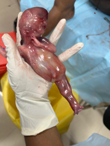
Figure 1: Face, neck and upper limbs were normal with bilateral legs fused and no external genitalia.

Figure 2: Posterior aspect showed a haemorrhagic swelling from occiput to lumbo- sacral area and there was a remnant attached to the anal area, anus absent.
Diagnosis of sirenomelia prenatally can be done by USG during the first trimester easily than the second trimester. This is because of the progressive decrease in liquor due to renal agenesis or dysplastic kidneys. A case series described 12 cases from various parts of the World and reported that 11/12 cases diagnosed between 11-13 weeks of pregnancy. They concluded that abnormal lower limbs or an intra-abdominal cyst located laterally during the first-trimester scan are the warning signs of sirenomelia [2].
Common associated anomalies with sirenomelia are absent external genitalia, ambiguous genitalia, imperforate anus, rectal atresia, absent urinary bladder, single umbilical artery, renal agenesis, esophageal atresia, omphalocele, pulmonary hypoplasia, cardiac defect, diaphragmatic hernia, lumbosacral/pelvic bone abnormities, and spina bifida. Even though Sirenomelia manifestation may overlap with caudal regression syndrome (CRS) and VACTERL association, they are different entities. Sirenomelia is classified into seven types based on skeletal structures of the lower limb [3]. In the present case there were no external genitalia and anus and there was vestigial remanant and no sacrum in addition to dysplastic kidneys. The haemorrhagic swelling on the back of this fetus may be due to vascular malformation. Sirenomelia is thought to be caused by a vitelline vascular steal phenomenon secondary to persistence of the vitelline artery. The single artery is formed by a coalescence of arteries that supply the yolk sac, arising from the descending aorta high in the abdominal cavity, and redirects blood flow from the developing caudal structures of the embryo to the placenta [4].
In ARKPD fetus on USG has enlarged kidneys with cysts of various sizes without corticomedullary differentiation and this disorder can be diagnosed in later gestational ages also. The differential diagnosis includes autosomal dominant polycystic kidney disease, Glomerulocystic cortical cysts, Hepatocyte nuclear factor-1-beta (HNF1B)-related cystic disease,Nephronophthisis and other ;multisystem disorders such as Meckel-Gruber syndrome, Bardet-Biedl syndrome, Joubert syndrome, and Jeune asphyxiating thoracic dystrophy etc. [5]. Genetic testing undertaken in this fetus keeping in mind ARPKPD, did not show any abnormalities. Chromosome and/or microarray genetic analyses undertaken in six cases of Sirenomelia by Sepulveda et al revealed normal karyotype with no copy number variants [2].
The roles of maternal diabetes, exposure to heavy metals, and radioactivity were described as important environmental risk factors. As it occurs sporadically in humans, it may be due to an autosomal-dominant genetic background or more likely due to a combination of genetic and environmental components [6]. A case report from North India revealed that MRI is necessary to accurately diagnose. By USG duodenal atresia and sacral dysgenesis were only made out by USG and kidneys and limbs were not visualised by this modality even at 26 weeks. MRI showed single hypoplastic lower limb and hypoplastic upper limb and the woman was a primigravida with occasional intake of Coccaine and alcohol [7].
Mermaid is a very rare and lethal congenital anomaly that can be easily missed by USG. High index of suspicion is necessary to diagnose by USG and it should be suspected in renal agenesis or when kidneys are visualised as polycystic or multicystic. When the limbs are straight and not moving or not seen separately or not visualised an MRI should be undertaken rather than chorionvillous biopsy. It is important to diagnose Sirenomelia early in gestation as it is a life threatening anomaly.
Copyright: © 2024 Dipali Singh and Paapa Dasari. This is an open-access article distributed under the terms of the Creative Commons Attribution License, which permits unrestricted use, distribution, and reproduction in any medium, provided the original author and source are credited.


 Open Access by
Acta Scientific is licensed under a Creative Commons Attribution 4.0 International License
Open Access by
Acta Scientific is licensed under a Creative Commons Attribution 4.0 International License
Based on a work at https://actascientific.com
ff