Rahul Deo Sharma1*, Anant Bangar2, Surendra Singh2 and Sonia Thakur2
1 Department of Pediatric Surgery, Ujala Cygnus Rainbow Hospital, Agra, India
2 Department of Pediatric Surgery, Lilavati Hospital & research Centre, Mumbai, India
*Corresponding Author: Rahul Deo Sharma, Department of Pediatric Surgery, Ujala Cygnus Rainbow Hospital, Agra, India.
Received: June 13, 2024; Published: June 30, 2024
Citation: Rahul Deo Sharma., et al. “Ewings Sarcoma Masquerading as an Infected Axillary Cyst”. Acta Scientific Paediatrics 7.7 (2024): 29-30.
Extraskeletal Ewing sarcoma (EES), a rare and aggressive malignancy within the Ewing sarcoma family of tumors (ESFT), can present diagnostic challenges due to its atypical locations and symptoms. Representing approximately 0.4 cases per million, EES commonly manifests as a rapidly growing, painful mass primarily in the soft tissues of the upper thigh, buttocks, upper arm, and shoulders, with common metastases in the lungs, bones, and bone marrow. This case report details an unusual presentation of EES mimicking a sebaceous cyst in a 10-year-old male. Initial symptoms included a progressively enlarging, painful axillary swelling with intermittent fever and redness. Ultrasound revealed a hypoechoic cystic lesion, and complete excision followed by histopathological analysis confirmed the diagnosis of Ewing sarcoma. Post-surgical management included chemotherapy and radiation therapy, leading to a favorable outcome with no disease recurrence at one year follow-up. The case emphasizes the critical importance of histopathological examination for accurate diagnosis and highlights the favorable prognosis of EES compared to its skeletal counterpart, particularly when diagnosed in younger patients”.
Keywords: Extraskeletal Ewing Sarcoma (EES); Ewing Sarcoma Family of Tumors (ESFT).
Extraskeletal Ewing sarcoma (EES) is a rare subtype within the Ewing sarcoma family of tumors (ESFT), characterized by small round cells with shared neural histology and genetic traits [1]. The ESFT spectrum includes classical Ewing sarcoma of bone (ESB), the second most common primary bone malignancy in children, peripheral primitive neuroectodermal tumor (pPNET), and Askin tumor of the chest wall. EES, identified in 1969, remains relatively undocumented with an incidence rate of 0.4 per million, significantly lower than ESB [1]. EES exhibits a bimodal age distribution, affecting individuals under 5 and over 35 years of age. This rapidly growing mass causes localized pain and typically arises in the soft tissues of the upper thigh, buttocks, upper arm, and shoulders. Common metastatic sites include the lungs, bones, and bone marrow, influencing the symptoms based on the primary and metastatic sites, which are present in 25% of cases at diagnosis.
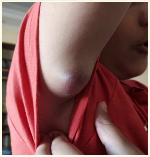
A 10-year-old male presented with a gradually enlarging swelling in the right axilla over three months, accompanied by intermittent pain, fever, and redness. There was no history of cough, cold, trauma, or weight loss. Physical examination revealed a 5x5 cm firm to cystic swelling in the dermal plane of the right axilla with prominent veins and tenderness. Systemic examination findings were normal. Blood tests indicated hemoglobin at 12.2 gm/ dl, TLC at 10220, platelets at 3.54 lakh, and serum creatinine at 0.38. Ultrasonography revealed an ovoid hypoechoic cystic lesion measuring 2.6x2.5x1.5 cm in the cutaneous and superficial subcutaneous plane of the right axilla. The infected swelling was completely excised and sent for histopathological examination, which confirmed a diagnosis of malignant Small Round Cell Tumor—Ewing’s sarcoma, with CD 99, Vimentin, and Cytokine d1 positivity. Post-diagnosis, a PET scan showed no active disease at the operative site, though there was low to moderate grade activity in minimally enlarged neck nodes suggestive of reactive nodes. The patient underwent another surgery for wide local excision, and histopathological examination showed no residual viable tumor. The patient began chemotherapy, receiving 17 cycles of vincristine and doxorubicin, followed by 25 cycles of 45Gy radiation therapy. The patient is currently asymptomatic with a one-year follow-up.

Figure 1
Extraskeletal Ewing’s sarcoma (EOES) is a malignant tumor of soft mesenchymal tissue, commonly affecting the lower extremities and paravertebral region. With an incidence rate of about 1.1% among malignant soft tissue tumors, EOES primarily affects individuals aged 5-25 years, showing a higher prevalence in males. The exact cause is unknown, but histologically, EOES is a neuroectodermal tumor. Hematoxylin-eosin staining reveals an intense blue color typical of ES, categorized under blue round cell tumors. Immunohistochemistry, crucial for differential diagnosis, often shows positive CD99/MIC2 markers in 98% of cases [3], though these markers can also be present in other blue round cell tumors like lymphoblastic lymphoma, rhabdomyosarcoma, small cell carcinoma, and poorly differentiated synovial sarcoma [2]. 30 However, in this case, other possibilities were excluded based on immunoreactive negativity to markers such as leukocyte common antigen (CD45/LCA), desmin, and broad-spectrum cytokeratin. Additional markers like epithelial membrane antigen (EMA), CD20/ L26, CD45-RO/UCHL-1, and terminal deoxynucleotidyl transferase (TDT) were also negative. Current treatment, as recommended by the National Comprehensive Cancer Network (NCCN), involves local treatment (surgery and/or radiotherapy) combined with chemotherapy, including alternating cycles of vincristine, doxorubicin, cyclophosphamide, and ifosfamide, etoposide every three weeks. EES generally has a better prognosis compared to skeletal Ewing’s sarcoma, with factors affecting prognosis being similar. Notably, localized EES has a higher five-year overall survival rate than localized skeletal Ewing’s sarcoma. Younger age at diagnosis (<14 years) is a strong predictor of favorable outcomes [4]. Recent reports indicate that patients with extraskeletal primary tumors tend to have better prognoses than those with skeletal primary tumors, with relapse rates as high as 30% in some series, leading to poor outcomes despite salvage chemotherapy attempts. Larger tumor size (>8 cm), metastatic disease at diagnosis, and relapsed disease are associated with worse outcomes.
This case of Extraskeletal Ewing’s sarcoma presenting as an axillary sebaceous cyst highlights the importance of histopathological examination of every excised lesion.
Copyright: © 2024 Rahul Deo Sharma., et al. This is an open-access article distributed under the terms of the Creative Commons Attribution License, which permits unrestricted use, distribution, and reproduction in any medium, provided the original author and source are credited.
ff
© 2024 Acta Scientific, All rights reserved.