Vinaykumar P Hedaginal1*, Shreya Bhate2, Fehmida Najmuddin3, Soumya Ahuja1 and Vaibhavi Pandya1
1 Junior Resident, Department of Paediatrics, DY Patil Deemed to be University, Navi Mumbai, India
2 Senior Resident, Department of Paediatrics, DY Patil Deemed to be University, Navi Mumbai, India
3 Associate Professor, Department of Paediatrics, DY Patil Deemed to be University, Navi Mumbai, India
*Corresponding Author: Vinaykumar P Hedaginal, Junior Resident, Department of Paediatrics, DY Patil Deemed to be University, Navi Mumbai, India.
Received: February 24, 2023; Published: April 04, 2023
Citation: Vinaykumar P Hedaginal., et al. “A Rare Case of Ambiguous Genitalia: Pseudovaginal Perineoscrotal Hypospidia”. Acta Scientific Paediatrics 6.5 (2023): 03-05.
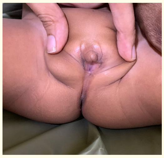
A 1 year old infant who was raised as female presented with microphallus, bifid labia like scrotum with bilaterally undescended testis, a blind ending vagina. There was a severe abnormal external genital development. But normal development of Wolffian duct derivatives including spermatic cord and testis with absent uterus and ovaries. Endocrinological investigations were within normal limits along with testosterone. Karyotyping and Whole exon sequence aided in diagnosing the patient with Pseudo-vaginal perineoscrotal hypospadias who was counselled and referred to higher centre for further management.
Keywords: Ambiguous Genitalia; Androgen Action; Dihydrotestosterone; Disorders of Sexual Differentiation (DSD), Genetic Testing.
Ambiguous genitalia is one of the rare problem encountered in newborns. It has a prevalence of 1 in 4500 live births. Only 20-40 % times the diagnosis of Disorder of Sexual development can be made with the help of recent advanced technology like Karyotyping, Genetic work up, an assay of hormones etc. Pseudo-vaginal perineo-scrotal hypospadias is one of the important cause of ambiguous genitalia in children caused by 5 alpha-reductase type 2 (5alpha-RD2) deficiency. The phenotype in these children can vary from underdeveloped male genitalia to fully developed female genitalia [1,2]. In this syndrome, the child who is genetically males contain normal male internal genitalia including testes but exhibit ambiguous or female external genitalia at birth. Here is a story of 1year old child who was raised as female was diagnosed to be a case of Pseudovaginal perineoscrotal hypospadias.
1 year old child was admitted in view of swelling on labia majora, on examination child was vitally stable with bifid scrotum like labia which contains palpable testis, 2 orifices visualised (anal and vaginal). USG abdomen was done which was showing oval hypoechoic testes like structures bilateral in inguinal region with attachment of spermatic cord in the proximal aspect right side 1.4cm x 0.6cm, left side 1.2cm x 0.5cm and? penis like structure is noted arriving from the root of pubic symphysis extending into the inter labia region. Uterine bud and ovaries are not visualised. Testosterone, FSH and LH levels were normal. Karyotyping was s/o 46 XY chromosomes indicating genotypically Male. Whole Exon genome was sent which showed Homozygous pathogenic variant SRD5A2 defect s/o Psuedovaginal perineoscrotal hypospadias. The child was referred to higher centre for Genetic specialist for further treatment.
Pseudovaginal perineoscrotal hypospadias is a autosomal recessive condition caused by 5 alpha reductase enzyme deficiency which converts testosterone to Dihydrotestosterone (DHT) . DHT or Testosterone binds to specific androgen receptors to form a complex that can regulate gene expression in majority of androgen target tissues [2].

Figure 1

Figure 2
Human embryo has the ability to differentiate into both male and female reproductive system. In the absence of Testosterone, female genital reproductive system is formed. SRY gene on short arm of Y chromosome initiates the differentiation of primordial undifferentiated gonadal ridge into male reproductive system by forming Testis which produces testosterone. Testosterone leads to formation of male internal genitalia including vas deferents, seminal vesicles and epididymis. DHT is required for formation of male external genitalia in Intrauterine life. Deficiency of % alpha reductase enzyme leads to low levels of DHT which intern lead to formation of ambiguous genitalia ranging from underdeveloped male genitalia to fully formed female external genitalia. So many parents rise their child as female [3,4].
In many cases during puberty due to low DHT and normal testosterone which will cause partial virilization and this testosterone is enough to cause some of the Male secondary sexual characters to develop including psychosexual behaviour, development of the embryonic wolffian duct, spermatogenesis, muscle development, voice deepening, and axillary and pubic hair growth. DHT is required for growth and development of Prostate, development of external genitalia and male patterns of body and facial hair growth [2].
The presentation can be varying in these children. Newborns might have female external genitalia with resemblance to labia Majora, which would be unfused labio-scrotal folds. The phallus may look like clitoris than a penis [5]. But the internal genitalia would be of males including seminal vesicles, vas deference, ejaculatory duct and epididymis. Testis may be present in inguinal sac or rarely abdominal cavity. So many children will be raised as females. At puberty they tend to develop male secondary sexual characters due to the effect of testosterone [3]. Phallus may get enlarged to form penis and the may present with deepened voices with facial beard and testis may descend to unfused labio-scrotal folds [5].
The 5 alpha reductase deficiency, partial androgen insensitivity syndrome and 17β-hydroxysteroid dehydrogenase type 3 enzyme deficiency are indistinguishable [6]. so proper evaluation with the help of recent advances like Karyotyping, Genetic work up, an assay of hormones etc. should be done to rule out the disease.
The treatment of a child depends on many factors, most important being gender of the child at the time of diagnosis. It would be better to raise the child as female if there is complete formation of female external genitalia. Later if the child wants to be female then corrective surgeries would be necessary along with removal of testis before the child attains puberty [7].
If the child is raised as a male, then corrective surgery should be done depending on the phenotype of the child and done during the first or second year of life [8]. Multidisciplinary, team including a geneticist, endocrinologist, paediatrician, paediatric surgeon, urologist, obstetrician, and gynaecologist help determine the appropriate course for the infant.
Copyright: © 2023 Vinaykumar P Hedaginal., et al. This is an open-access article distributed under the terms of the Creative Commons Attribution License, which permits unrestricted use, distribution, and reproduction in any medium, provided the original author and source are credited.
ff
© 2024 Acta Scientific, All rights reserved.