Metin Celalettin*
Victoria University, Institute of Telecommunications, Electronics, Photonics and Sensors, Melbourne, Australia
*Corresponding Author: Metin Celalettin, Victoria University, Institute of Telecommunications, Electronics, Photonics and Sensors, Melbourne, Australia.
Received: January 21, 2022; Published: January 20, 2023
Citation: Metin Celalettin. “A Paediatric Magnetic Resonance Imaging Technique Substituting Fluid-Attenuated Inversion Recovery Time of the Ernst Angle on Protons Within Paediatric Anatomy to Removing the Need to Administer an Anaesthetic Newborns, A Short Communication”. Acta Scientific Paediatrics 6.2 (2023): 21-22.
Within the boundaries of extant Magnetic Resonance Imaging (MRI) technology for paediatric patients, after 30 days of age, an intravenous sedative is administered. MRI is a non-invasive imaging procedure that uses a powerful magnetic field and radiofrequencies and records neuro-anatomical structures at an anatomical target location. Imaging anatomical abnormal and normal tissues in specific cases necessitate accepting the risk of causing harm to an infant. This article explores the benefits of an MRI using an alternative to the imaging protocol [1,11].
Keywords: Fluid-Attenuated Paediatric; Anatomy; Newborns.
The risk is in administering the sedative, or a general anaesthesia. This is due to the need for the patient to remain still. The risk of a paediatric fatal event under anaesthesia is one in 300,000 per annum. While this a relatively low risk in itself, the extant conjecture annual growth rate (CAGR) is of concern [1]. Paediatric patients are on a significant incline because of the procedure becoming available to second and third world countries, more governments are subsidizing MRI procedures, and the value of MRI imaging over all other imaging techniques [1,2,4,7].
This article considers the ethics and plausible biomechanical solutions is in proposing a paediatric MRI capability that removes the need to administer a general anaesthesia to a newborn. Originally designed to protect stealth fighter jets from quantum radar detection, this technology could be utilized as a safer means to medically image newborns [3,11]. The third part of this series of short-communications details both the protocol and changes that focus on a significantly more advanced method of measuring the fluid-attenuated inversion recovery time.
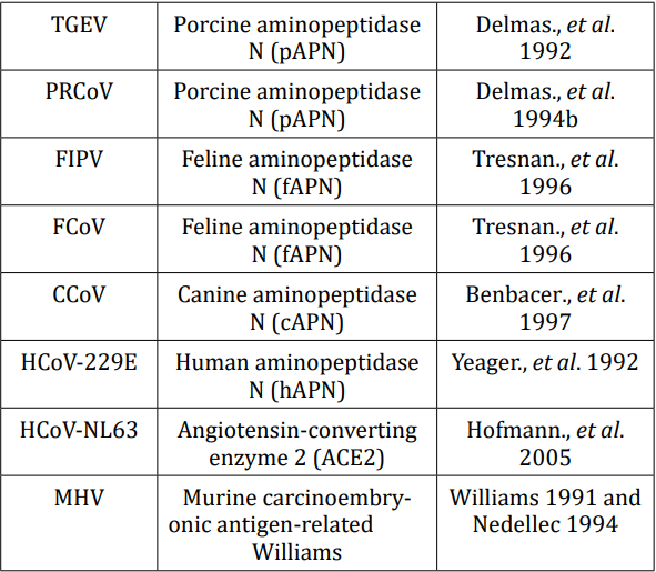
The receptors in Table 1 are those for severe acute respiratory syndrome-2, (SARS-2) each one a separate molecular compound with charges attributed able to their molecular structures [12].

Table 1: Cellular receptors for coronaviruses.
The difference between the polarization of human tissues during an MRI is to differentiate between the Ernst angle of spin polarized protons and measure extant fluid attenuation inversion recovery times. This requires a newborn to be administered anaesthesia during imaging to keep them still so as to repeat the procedure as required under extant technological limitations [1,3,10-12].
The brain is surrounded by cerebrospinal fluid (CSF), which has roughly the same signal intensity on images as brain tissue. Pulse sequences makes hyperintense signals from water, turn hypointense (black) on T2 images while keeping lesions hyperintense and recognizable. This is consistent with other tissues in the body; their FLAIR signals are too similar for a single quarter-second image [57,9].
In measuring the fluid-attenuated inversion recovery time of the Ernst angle on protons within the human anatomy, several measurements are required, necessitating a radically more accurate technique.
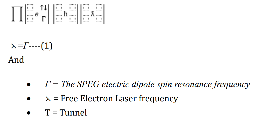
‘Celalettin’s 1st Paradigm’ expresses the behaviour of SARS-2 described by

‘Celalettin’s 1st Paradigm’ at Equation 1, can be used to measure the electric charges of the SARS-2 molecular constructs.
The paediatric ‘Hand-Held Magnetic Resonance Imaging Device’ (H2MRI) © in Part 2 and 3 of this Short Communication will further dissect and manipulate the paradigm in order to focus on the way in which extant MRI machines measure polarized Ernst angles on protons within anatomy. This could be engineered into a HandHeld Magnetic Resonance Imaging Device (H2-MRI©), which would remove the need to administer a sedative to newborns in addition to providing a more accurate image.
Copyright: © 2023 Metin Celalettin. This is an open-access article distributed under the terms of the Creative Commons Attribution License, which permits unrestricted use, distribution, and reproduction in any medium, provided the original author and source are credited.
ff
© 2024 Acta Scientific, All rights reserved.