A Badre*, T Faid, M Lehlimi, M Chemsi, A Habzi and S Benomar
Neonatal Medicine and Resuscitation Department, Abderrahim Harouchi Mother and Children's Hospital, Ibn Rochd University Hospital, Casablanca, Morocco
*Corresponding Author: Sangita D Kamath, Consultant, Department of Medicine, Tata Main Hospital, India.
Received: August 27, 2021; Published: October 16, 2021
Citation: A Badre., et al. “Mentosternal Dysraphia: A Case Report”. Acta Scientific Paediatrics 4.11 (2021): 30-32.
Mentosternal dysraphia is a congenital abnormality of the anterior part of the neck. It is rare and little known, and accounts for less than 2% of all cervical congenital malformations. The embryological origin is not fully elucidated. The embryologic origin is not fully understood and is thought to be the consequence of a defect in the median mesodermal fusion of the first and second gill arches between the 3rd and 4th week of embryonic development. It corresponds to strained flanges between the chin and the sternum. Its diagnosis is clinical and no further examination is necessary. The treatment is surgical and must be done during the first months of life to avoid mechanical and infectious complications mainly. Pediatricians, dermatologists and ENT specialists must recognize these lesions at an early stage to allow early and appropriate management. We report the observation of a newborn with mento-sternal dysgraphia diagnosed and operated on in our health establishment. The objective of this work is to study the clinical aspect, the modalities of therapeutic management and the evolution of this exceptional congenital malformation.
Keywords: Congenital Malformation; Mento-Sternal Dysgraphia
Mentosternal dysraphia is a rare congenital anomaly of the anterior part of the neck. The embryological origin is not completely understood. It would be the consequence of a defect of median mesodermal fusion of the first and second branchial arches between the 3rd and the 4th week of embryonic development [1]. This anomaly corresponds to straps stretched between the chin and the sternum. Its diagnosis is clinical and no additional examination is necessary. Management is surgical and must be performed during the first months of life to avoid mechanical complications mainly represented by retromandibular, but also infectious complications [2].
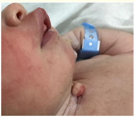
We report a case of mentosternal dysraphia diagnosed at birth in the Lalla Meryem maternity unit of the A. Harrouchi Mother and Children's Hospital. The newborn male R.B., was issued from a poorly monitored pregnancy, brought to term, with the notion of 3rd degree consanguinity. The delivery was performed by caesarean section for pre-eclampsia with good adaptation to extrauterine life, the Apgar score at the 1st minute being 10/10. Clinical examination found a pink, responsive, good-toned, hemodynamically and respiratory stable newborn, with a respiratory rate of 48 cpm, 98% oxygen saturation in the open air, a heart rate of 126 bpm, a cutaneous repolarization time less than 3 seconds. Archaic reflexes were present. We note at the anterior and middle part of the neck, 2 cm above the sternal fork, a formation measuring 3 x 1 cm with a large vertical axis, corresponding to a vertical bead with an excrescence of cartilage consistency (Figure 1). In hyperextension of the head, palpation of the neck notes a subcutaneous band extending from the mandible to the upper edge of the sternal fork. In addi to the upper edge of the sternal fork. In addition, a weeping inferior fistula and retrognatism were noted in our patient (Figure 2). The rest of the somatic examination does not reveal any abnormality and the malformative assessment is negative. A clinical follow-up was planned until the age of 6 months, in concertation with the pediatric surgeon, and then a surgical treatment with a Z-plasty was realized under general anesthesia with simples post-operatory suites (Figure 3). The anatomopathological study of the operative part confirms the diagnosis of mento-sternal dysraphy.

Figure 1: Mentosternal dysraphia, head in hyperflexion.

Figure 2: Mentosternal dysraphia, lateral view; Retrognatism.

Figure 3: The operative part.
Mental-sternal dysraphia is a rare and little-known birth defect. It constitutes less than 2% of all cervical congenital malformations [3]. These malformations are generally sporadic and no familial case has been described [4]. The pathogenesis would seem to be a defect of mesodermization of the arches by quantitative or qualitative deficit of the cells resulting from the neural crests which would be at the origin of a disorder of the mesodermal coalescence of the first and second right and left branchial arches on the ventral line [5]. Several hypotheses have been advanced to explain the mechanism of this incomplete fusion of the branchial arches [5]. Some authors have reported that abnormalities in the course of cell migration, a quantitative deficit of cells originating from neural crests or abnormalities in the interaction of these cells with the surrounding environment may be the cause of a fusion defect [6].
The diagnosis of this malformation is purely clinical [1]. It is most often done at birth, in the first days or weeks of life. The dysraphia extends between the chin and the sternal fork and is made of a vertical fibrous tract which descends from each side of the chin symphysis towards the sternum. On this route, skin buds are often found. A fibrous mass overcomes dysraphia and may end in a fistulous cul-de-sac [7]. It is important to note that there is a great variability of clinical presentations. The lesion can be present at any level between the mandible and the sternal manubrium. Its size can be very variable [8].
In our patient, the mento-sternal flange was the predominant element with a median excrescence of cartilage consistency associated with an inferior weeping fistula. We also note the existence of retrognatism (Figure 1 and 2).
In the vast majority of cases, this malformation is isolated. However, few cases of association with other malformations have been reported, including a cleft of the tongue, lower lip or mandible [4].
No additional examination or malformative assessment are necessary in classic forms [9]. If the fistula involves the lower lip or the mandible, a work-up for a heart defect is recommended as well as a cervical ultrasound to check the thyroid, the cartilage of the larynx for possible cysts [10].
The treatment is surgical. Surgery is formal but not urgent. It must be performed in the first months of life to avoid functional and morphological disorders of the neck and mandible as well as infectious complications [2]. It will be performed under general anesthesia and consists of a complete removal of scar tissue and fistulas, supplemented by one or more “Z” plasties to prevent the occurrence of a scar in the neck [11]. It should be avoided during the period of teething as hypersealorrhea could compromise the quality of healing [4]. Rigorous long-term monitoring is necessary because of the frequency of postoperative recurrence of fibrous retraction [6].
In our case, a clinical follow-up was planned until the age of 6 months, at the requested of the pediatric surgeon, and then a surgical treatment with a Z-plasty was realized under general anesthesia with simples post-operatory suites.
Mentosternal dysraphia is one of the rarest congenital malformations of the neck. The diagnosis is clinically obvious and does not require imaging, but it is often unrecognized at birth. Pediatricians, pediatric surgeons, otolaryngologists and dermatologists must recognize this malformation early to allow appropriate management in order to improve neck mobility and reduce the risk for long-term cervical tethering and mandibular defects.
Copyright: © 2021 A Badre., et al. This is an open-access article distributed under the terms of the Creative Commons Attribution License, which permits unrestricted use, distribution, and reproduction in any medium, provided the original author and source are credited.
ff
© 2024 Acta Scientific, All rights reserved.