Edwin Dias1*, Shwetha P N2, Vinod Prem Singh3 and Kiran Raj H4
1Professor and HOD, Department of Pediatrics, Srinivas Institute of Medical Science and Research Centre, Mangalore, India
2Junior Resident, Department of Pediatrics, Srinivas Institute of Medical Science and Research Centre, Mangalore, India
3Professor, Department of Pediatric Surgery, Srinivas Institute of Medical Science and Research Centre, Mangalore, India
4Assistant Professor, Department of Pediatrics, Srinivas Institute of Medical Science and Research Centre, Mangalore, India
*Corresponding Author: Edwin Dias, Professor and HOD, Department of Pediatrics, Srinivas Institute of Medical Science and Research Centre, Mangalore, India.
Received: April 06, 2021; Published: April 19, 2021
Citation: Edwin Dias., et al. “A Case Report of Meconium Plug Syndrome with Intestinal Obstruction in a SGA, Preterm 34 Week Gestation Baby”. Acta Scientific Paediatrics 4.5 (2021): 29-33.
Introduction: Meconium plug syndrome is most commonly seen in sick, premature infants and those having small left colon. Incidence of meconium plug syndrome ranges from 0.5-1 case per 500 live-births. Clinically, presents with inability to pass meconium, distended abdomen, vomiting.
Objective: To report a case of meconium plug syndrome in a Small for Gestational Age (SGA), preterm baby.
Clinical Case: The case to be presented is of a SGA, preterm boy baby who had presented at 24 hours of life with abdominal distension and not passed meconium since birth. Abdominal x-ray suggestive of dilated bowel loops with no air fluid levels.
Treated by suppository and rectal thermometer passed cautiously at 72 hours of life, following which thick gelatinous mucoid material -meconium plug was expelled out.
On day 4 of life, baby still did not pass meconium. Hence, nasogastric tube was inserted per rectally and bowel wash was given. About 30ml of meconium was aspirated. Abdominal distension gradually reduced. On day 5 of life, baby passed about 5ml of wellformed meconium followed by normal passage of meconium.
Conclusion: Meconium plug can be relieved by glycerin suppository. Rectal irrigation with pre-warmed 0.9% normal saline may also help in relieving meconium plug. Contrast enema helps in delineating the meconium plug as well as to relieve obstruction, therefore acts as both diagnostic and therapeutic tool. The overall outcome of the baby for the treatment provided was positive and child improved symptomatically.
Keywords: Meconium Plug Syndrome; Hirschsprung’s Disease; Cystic Fibrosis; Glycerin Suppository; Contrast Enema
The term “Meconium plug syndrome” was initially described by Clatworthy., et al. in 1956 [1]. In the past, the hypothesis of altered colonic motility or altered character of meconium causing mechanical obstruction was made. The term “plugged-up babies” was used to describe this condition of meconium plug syndrome. Clinical features include inability to pass meconium, vomiting, and abdominal distension. Abdominal x-ray shows features of intestinal obstruction. The meconium plug may sometimes get dislodged after stimulation of the anus and rectum. Most often, colon will be functionally unaffected. Historically, causes of meconium plug syndrome have been thought to include cystic fibrosis, small left colon syndrome, Hirschsprung disease, magnesium tocolysis for preeclampsia. Other causes are congenital hypothyroidism, maternal opiate use and neuronal intestinal dysplasia. Following resolution of a meconium plug, a sweat chloride test should be done to rule out cystic fibrosis (CF) and a thyroid stimulating hormone (TSH) level estimation to exclude congenital hypothyroidism. A rectal biopsy should be performed to evaluate for Hirschprung’s disease, if there is a dysfunctional stooling pattern even after resolution of the plug [2].
Meconium plug syndrome has a wide range of severity, starting from meconium plug syndrome to the meconium peritonitis.
We report a case of meconium plug syndrome with intestinal obstruction in a SGA, preterm baby and review the clinical presentation, radiological features and treatment of this condition.
A SGA, preterm boy baby was delivered via emergency cesarean section, indication being preterm with non-reassuring fetal heart rate and maternal gestational hypertension. Baby was born to a 25-year-old primigravida mother at 34 weeks period of gestation with birth weight of 1.63kg. She was booked case, had regular antenatal check-up, had taken iron, calcium and folic acid supplements during pregnancy. Mother was diagnosed with gestational hypertension since 5th month of pregnancy and was on antihypertensive medication (Nifedipine).
Baby was kept nil per oral since birth in view of respiratory distress and was started on IV fluids, oxygen through nasal prongs and empiric antibiotic coverage through ampicillin and gentamicin. Umbilical venous catheterization was done. Nasogastric tube (NG tube) was inserted to decompress the stomach. At 24 hours of life, mild abdominal distension was noted with dilated abdominal veins, sluggish bowel sounds. Anal opening was patent. Abdominal Girth was 26cm, increase in AG compared to baseline. Chest and abdominal x ray was taken (Figure 2). Abdominal x-ray showed dilated bowel loops with no air fluid levels. NG tube aspiration was continued every two hourly.
At 6 hours of life, septic work up was done which showed Hb: 18.2g/dl, PCV: 55.7%, WBC: 14,570cells/cumm, lymphocytes: 14%, neutrophils: 75%, eosinophils: 2%, Basophils and monocytes: 0%, platelet count: 1.41lakhs, peripheral blood smear showed macrocytic blood picture with I: T Ratio of 0.1, CRP: negative, S. calcium: 11.05mg/dl.
At 44 hours of life, as baby did not pass meconium, pediatric surgeon reference was sought. On examination, abdomen was distended, skin over abdomen was stretched and shiny (Figure 1), dilated bowel loops and peristaltic movements were visible. AG increased to 27.5cm. Tympanic note heard on percussion. Sluggish Bowel sounds heard on auscultation. Removed umbilical venous catheterisation and antibiotics were stepped up as blood culture reports showed klebsiella which was sensitive to piperacillin and tazobactam. Ultrasound abdomen revealed prominent and oedematous bowel loops with hyper-echoic foci. Stool for occult blood was negative.
At 72 hours of life, suppository chip was put which got melted, then rectal thermometer was passed cautiously. There was no explosion of flatus or meconium following insertion of rectal thermometer. Hence, Hirschsprung’s disease was ruled out. Following which after about 4 hours, thick gelatinous mucoid material –meconium plug (Figure 3) was expelled out.
On day 4 of life, baby didn’t pass meconium, abdominal distension increased to 28cm. Cautiously, No: 8 NG tube was inserted per rectally and bowel wash was given with pre-warmed 0.9% Normal saline. Approximately, 30ml of meconium was aspirated. Following which abdominal distension gradually reduced from 28cm to 26cm. Later, NG feeds started with 2ml every two hourly and increased as baby tolerated.
On day 5 of life, baby passed about 5ml of well-formed meconium. Following which the baby had normal meconium output.
On day 6, NG tube removed as baby was tolerating palladai feeds. Later, direct breast feeds started. Baby tolerated the feeds and bowel output was normal (Figure 4). The diagnosis of meconium plug syndrome leading to intestinal obstruction was made retrospectively.
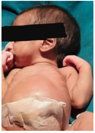
Figure 1: Abdomen is distended, skin over abdomen is stretched & shiny, dilated bowel loops, visible peristaltic movement.
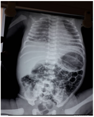
Figure 2: X-ray suggestive of distended bowel loops with no air fluid levels.
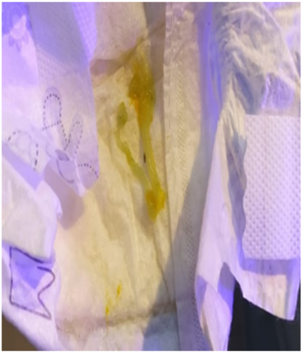
Figure 3: Thick, inspissated mucoid material –meconium plug
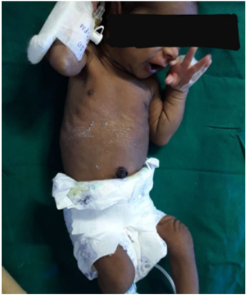
Figure 4: Post procedure - active baby, abdominal distension subsided.
Meconium plug syndrome clinically presents like other distal intestinal obstruction. Main clinical feature will be inability to pass meconium within 24 to 48 hours of life [3].
Inspissated meconium within the lumen of the intestine causes the mechanical obstruction. This will be seen on abdominal x-rays as mottled densities or bulky intraluminal colonic masses. If the plain x-ray does not readily show features of meconium plug, colonic contrast enema may be used which shows the typical “tape or ribbon-like or sausage shaped defects” in the colon. Sometimes, it can be even an incidental finding in newborns when they does not show any signs of intestinal obstruction clinically [4]. Complications of meconium plug syndrome include mucosal ulceration and intestinal perforation. After relieving the meconium plug, newborn should be followed up for any signs of congenital aganglionic megacolon [5].
Prompt recognition along with an early and aggressive conservative management is essential to prevent complications and spare surgical interventions. Gastrografin enema is proved to be safe and effective if promptly implemented in a suitable environment and by experienced hands [6]. Presence of a meconium plug on contrast enema most often correlates with a benign clinical course. If neonate is having abnormal stooling pattern, further evaluation is indicated. When meconium plug syndrome is identified as pellets and present only in the colon, then a diagnosis of cystic fibrosis may be a rare event [7].
Meconium plug syndrome is usually benign. It is due to immaturity of the colonic ganglion cells. Plain roentgenograms in these neonates with meconium plug syndrome show multiple distended bowel loops and other features of lower intestinal obstruction. On a contrast enema study, multiple filling defects (i.e., meconium plugs) may be seen, although the amount of colonic meconium is variable and discrete meconium plugs may not be present. Despite characteristic specific pattern on contrast enema depicting functional immaturity of the colon, an important differential consideration is Hirschsprung disease. Unusual cases of Hirschsprung disease that involve a long segment and have a transition point at the splenic flexure may completely mimic functional immaturity of the colon on imaging studies. Therefore, neonates with a presumed diagnosis of functional immaturity of the colon whose symptoms do not resolve after therapeutic enema should be considered for colonic biopsy to make a definitive diagnosis [8].
In newborn with features like meconium ileus, laparotomy followed by ileostomy is the line of management. Once the obstruction due to meconium plug is relieved, intestinal activity improves and oral feeding has to be promoted. Pediatric surgery consultation is advocated [9].
It was hypothesized that meconium plug syndrome is caused due to muscular weakness and colon immaturity. This plug causes obstruction and leads to development of clinical features. Clinically, the most significant features are the onset of obstructive symptomatology and a failure to pass normal meconium within the first 30 hours of life. The rectal examination is usually normal, but occasionally the plug may be palpable [10].
Retained meconium will be visualized as multiple filling defects in the left colon on contrast enema. In meconium plug syndrome, the rectum will be of normal caliber. Enema dislodges meconium plug either during or after the procedure, thereby helps in both diagnosis and treatment [11].
Repeated enemas and surgical decompression is rarely required. There is association of meconium plug syndrome with Hirschprung disease and cystic fibrosis. Hence, diagnostic tests are conducted to rule out their presence [12]. In a study conducted by Alex., et al. total sample size was 61 cases of meconium plug syndrome. Among which, about 30% cases had spontaneous resolution. Only two patients underwent surgery due to peritonitis and progressive distension. It concluded that according to survey conducted, association of magnesium tocolysis seen in 16% of cases and Hirschprung’s disease in 3.2% of cases [13].
Incidence of meconium plug syndrome is equal in both sexes. Risk factors for meconium plug syndrome includes preterm infants with small left colon, infants of diabetic mothers, and mothers who were on tocolytics. Meconium plug syndrome is associated with Hirschsprung disease and cystic fibrosis to certain extent [14]. Sweat chloride test has to be done to rule out cystic fibrosis [15]. If rectal biopsy depict the presence of ganglion cells, then Hirschsprung disease will be ruled out [16].
Intestinal obstruction accounts for about 33% of all Neonatal Intensive Care Unit (NICU) admissions. Inability to pass meconium within first 30 hours of life in newborn, intolerance to feeds, distended abdomen, biliary vomiting are characteristic features of intestinal obstruction and other diagnosis has to be considered based on anatomic, metabolic, and functional parameters. Newborns with meconium plug syndrome have good outcome, irrespective of period of gestation. Correlation of clinical and radiological findings is important to make a correct diagnosis, treat appropriately and to get a positive outcome.
None.
Copyright: © 2021 Edwin Dias., et al. This is an open-access article distributed under the terms of the Creative Commons Attribution License, which permits unrestricted use, distribution, and reproduction in any medium, provided the original author and source are credited.


 Open Access by
Acta Scientific is licensed under a Creative Commons Attribution 4.0 International License
Open Access by
Acta Scientific is licensed under a Creative Commons Attribution 4.0 International License
Based on a work at https://actascientific.com
ff
ff
© 2024 Acta Scientific, All rights reserved.