Hazar Khankan*
Pulmonology Division, Department of Pediatrics, Children's Damascus University Hospital, Syria
*Corresponding Author: Hazar Khankan, Pulmonology Division, Department of Pediatrics, Children's Damascus University Hospital, Syria.
Received: October 16, 2019; Published: October 29, 2019
Citation: Hazar Khankan. “Acute Chest Syndrome in A 10-Year-Old Male with Sickle Cell Disease ”. Acta Scientific Paediatrics 2.11 (2019):65.
A 10-year-old male with known sickle cell disease was admitted to Children's Damascus University Hospital with a history of tightness, shortness of breath, severe chest pain, and back pain for 2 days.
On physical examination, he had crackles with slightly diminished breath sounds on the base of both sides of the chest. Oxygen saturation was 94% on room air. The patient was hemodynamically stable.
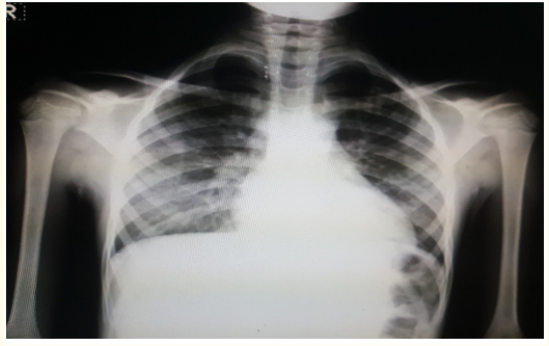
Investigations revealed hemoglobin of 8.4 mg/dl and total leucocyte count of 16,000/mm3 (Polymorphs 78%, lymphocytes 16%). CXR showed lateral opacity in the right mid- and lower- lung field and lateral opacity in the left mid-lung field (Figure 1). Chest computed tomography images showed bilateral consolidations predominating at lung bases (Figure 2,3).

Figure 1

Figure 2

Figure 3
A diagnosis of acute chest syndrome (ACS) was made and the management included analgesics, intravenous fluids, oxygen, wide spectrum antibiotics and red blood cell transfusion.
Copyright: © 2019 Hazar Khankan. This is an open-access article distributed under the terms of the Creative Commons Attribution License, which permits unrestricted use, distribution, and reproduction in any medium, provided the original author and source are credited.
ff
© 2024 Acta Scientific, All rights reserved.