Lee KJ*, Theenesh B and Ibrahim MI
Foot and Ankle Unit, Orthopaedic Department, Hospital Raja Permaisuri Bainun Ipoh, Malaysia
*Corresponding Author: Lee KJ, Foot and Ankle Unit, Orthopaedic Department, Hospital Raja Permaisuri Bainun Ipoh, Malaysia.
Received: August 30, 2024; Published: September 20, 2024
Citation: Lee KJ., et al. “Reliability of Radiological Parameters in Assessment of Haglund’s Syndrome in Malaysian Patients”. Acta Scientific Orthopaedics 7.10 (2024): 08-12.
Background: Haglund’s deformity is a significant cause of Haglund’s syndrome. Many radiological parameters have been used to define its presence. Commonly used parameters include the Fowler-Philip angle, calcaneal pitch angle, parallel pitch lines and the X/Y ratio. This paper seeks to apply these radiological parameters to a Malaysian cohort of patients to assess its reliability.
Methods: This is a single centre study with 21 symptomatic and 21 asymptomatic patients. Weight bearing lateral x rays were obtained and measurement of radiological parameters were performed independently by 2 Foot and Ankle surgeons. The parameters measured were the Fowler-Philip angle, calcaneal pitch angle, parallel pitch lines and the X/Y ratio.
Results: All the measured radiological parameters did not show high specificity and sensitivity. There were no statistically significant differences for measurements between the symptomatic and asymptomatic group. Both the groups had positive radiological parameters that suggested Haglund’s deformity.
Discussion: Each radiological parameter had an inherent weakness that could not be addressed. The Fowler-Philip angle does not take into account calcaneal verticality or length, the calcaneal pitch does not take into account the calcaneal morphology, parallel pitch lines do not consider the location of the superior projection and the X/Y ratio does not take into account the verticality of the calcaneum.
Conclusion: There is no gold standard radiological parameter for assessment of symptomatic Haglund’s deformity. The parameters tested showed poor sensitivity and specificity.
Keywords: Haglund’s; Fowler-Philip Angle; Parallel Pitch Lines; X/Y Ratio
Posterior heel pain is a common problem affecting up to 13% of adults over the age of 50 [1]. Pain is often attributed to insertional Achilles tendinopathy, retrocalcaneal bursitis or Haglund deformity. Often, these conditions exist together and are collectively known as Haglund syndrome [2]. The causes of Haglund syndrome are varied. The Haglund deformity, a bony prominence of the posterosuperior aspect of the calcaneus, was first described by Patrick Haglund in 1928 [3]. It has been theorised that this prominence causes impingement of the posterior part of the greater tuberosity of the calcaneus and neighbouring soft tissue: precalcaneal bursa, Achilles tendon, retrocalcaneal bursa and the subcutaneous tissue [4]. The Haglund deformity is oft mentioned in literature and is still a commonly debated topic as there is no standardised radiological measurement of the deformity.
Multiple radiological indices have been described to measure this deformity such as Fowler-Philip angle [5], calcaneal pitch angle [6], Chauveaux-Liet angle [7], parallel pitch lines(8) and the X-Y ratio [4a]. The X-Y ratio is of particular interest as the original description by Haglund in 1928 was of excessive heel length but there was no radiological proof provided at that time [3].
Studies to validate these parameters have been performed and show varying degrees of specificity and reliability. This may account for the varying methods and results of surgical treatment for Haglund’s syndrome [9-12].
The purpose of this paper is to determine if these most frequently used radiological indices are reliable methods of assessment of symptomatic Haglund’s deformity in the setting of Malaysian, and by extension, Asian patients.
This is a single-centre retrospective observational study which included 21 patients with Haglund’s syndrome requiring surgical treatment, and a control group of 21 patients who came for consultation of other foot and ankle pathologies without any hind foot complaints.
The Haglund group consists of 11 female and 10 male patients, with 10 patients having left sided calcaneum and 11 patients having right sided calcaneum pathology. The mean age of patients was 47 years (range 25-70). Inclusion criteria comprised of posterior heel pain with a calcaneal hump, evidence of irritation of skin by shoewear posterior strip, calcaneal insertion pain and tenderness, painful pre- or retro-calcaneal bursitis, radiologic evidence of enthesis calcification and enthesophytes.
The control group consists of 7 female and 14 male patients, with 10 left sided and 11 right sided calcaneums investigated. These patients have a mean age of 42 years (range 23-74). They had no symptoms or signs of hind foot pathology and were consulted for forefoot and ankle pathology.
The exclusion criteria applied were any calcaneal tendon body tendinopathy, neuromuscular conditions, inflammatory rheumatism, and any history of hindfoot trauma or surgery.
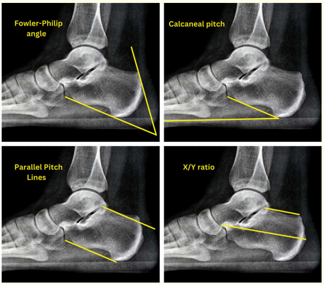
All patients had weight-bearing x rays of the foot and ankle which comprised of an AP and strict lateral view. Standard radiological criteria generally agreed in literature was used to quantify the bony abnormality seen. Measurements were taken on the lateral weight bearing views. The criteria of each measurement is as follows and illustrated in Figure 1

Figure 1: Radiological Indices for measurement of Haglund’s deformity.
The measurements were recorded independently by 2 Foot and Ankle surgeons (IMI, TB) and results were only made known to the principal researcher (LKJ). Inter-observer reliability was assessed with kappa coefficient and t-test was used to compare the means of the 2 X/Y ratio measurements made by the observers. Statistical analysis was performed using SPSS Statistics software V26 (IBM Corp., Armonk, NY). The significance threshold was set at p < 0.05.

Table 1: Radiologic measurement results.
Table 1 shows the radiological measurements obtained for the symptomatic and asymptomatic groups. In the Haglund group, an abnormal Fowler-Philip angle was only measured in 1 patient (79.4), with the rest being normal (mean 56.2 ± 9.2, range 44.179.4). Calcaneal pitch was pathologic in 14.3% of cases, with a mean of 27.1 ± 5.1 (range 19.5-39.6). Parallel pitch lines test was positive in only 13 patients (61.9%). X/Y ratio was positive in 57% of patients with a mean of 2.6 ± 0.4 (range 2.2-3.8).
In the asymptomatic group, Fowler-Philip angle was normal in all patients with a mean of 57.4 ± 6.9 (range 43.9-69.7). Calcaneal pitch was pathological in 19% of cases, with a mean of 24.6 ± 8.1 (range 11.8-40.7). Parallel pitch lines test was positive in 42.9% of cases (9). X/Y ratio was positive in 33% of patients with a mean of 2.6 ± 0.2 (range 2.2-3). There was no statistically significant difference noted between the groups for all measurements obtained.
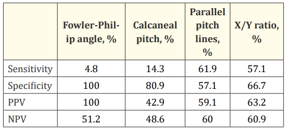
Table 2 shows the sensitivity, specificity, positive predictive value (PPV) and negative predictive value (NPV) of the measurements obtained. None of the radiological indices had a high sensitivity and specificity value.

Table 2: Sensitivity, specificity, PPV, NPV.
Posterior heel pain is a common problem, and the ‘pump bump’ is seen as a significant contributor to its aetiology. The description and classification of the Haglund deformity has led to an array of radiological studies which seek to define its existence consistently. Early reports of every proposed radiological parameter have shown promising results, but inconsistencies in study design and failure to reproduce similar positive correlation in subsequent studies have led to a failure of adoption of any single measurement as a gold standard in diagnosis. In this study, none of the radiological parameters showed high sensitivity or specificity.
Part of the reason that there is no universally accepted radiological parameter is due to the inherent weakness of each different measurement. The original paper by Fowler in 1945 showed the normal range of the Fowler-Philip angle to be 44 to 69 degrees, yet the diagnostic threshold was set at 75 degrees with no clarification as to its basis. Further studies have shown that symptomatic Haglund patients rarely had an Fowler-Philip angle of over 75 degrees. The weakness of the Fowler-Philip angle is also that it does not account for the verticality of the calcaneum. In a cavus foot, the Haglund projection is more prominent posteriorly even with a normal Fowler-Philip angle. In this study, there was only one patient who had a Fowler-Philip angle of >75 degrees and symptomatic patients had an angle as low as 44.1 degrees.
The parallel pitch lines do not take into account the position of the superior projection. Projections which are situated more anteriorly would not be causing any impingement or tension over the posterior structures. Moreover, it only takes into account the vertical height and neither the inclination nor length of the calcaneum, which may be a greater source of pressure on the Achilles. In this study, 61.9% of the symptomatic and 42.9% of the asymptomatic group were classified to have a positive parallel pitch line test.
The calcaneal pitch angle serves to illustrate the verticality of the calcaneum. In a more vertical calcaneum, any posterosuperior projection would be more prominent and effect the posterior structures more significantly. The change in position may also result in extra traction on the Achilles tendon. Bulstra., et al. [13] found a correlation between calcaneal verticality and Haglund’s syndrome, however the subgroup analysis found this to be significant only in female patients.
The X/Y ratio can serve as a guide in choosing between the 2 surgical treatments that modify the morphology of the calcaneum: a Zadek subtraction osteotomy or a resection of the posterosuperior corner (calcaneoplasty). An overly long calcaneum (X/Y <2.5) would benefit from the Zadek osteotomy whereas the short one may not. In this study however, there is no statistically significant benefit in the use of this parameter for diagnosis but it may serve its intended purpose well as stated. The ratio was positive in 57% of symptomatic patients and 33% in the control group.
It can be summarised that the aetiology of Haglund’s syndrome is not singular, but multivariate, and no single radiological parameter has satisfactorily taken into account all the factors to determine a pathological calcaneum. A combination of a normal calcaneum with abnormal verticality may lead to symptoms, while an abnormal calcaneum can be asymptomatic if it lies in a more horizontal position. The clinician will do well to select the appropriate treatment or procedure available on a personalised basis and use the radiological parameters as an adjuvant in overall management.
Copyright: © 2024 Lee KJ., et al. This is an open-access article distributed under the terms of the Creative Commons Attribution License, which permits unrestricted use, distribution, and reproduction in any medium, provided the original author and source are credited.