Mohamed Abuzaid*
Departement of Gynaecology and Obstetrics, St. Vinzenz Hospital Hanau, Germany
*Corresponding Author: Mohamed Abuzaid, Departement of Gynaecology and Obstetrics, St. Vinzenz Hospital Hanau, Germany
Received:October 16, 2024;; Published: October 23, 2024
Citation: Mohamed Abuzaid. “Laparoscopic Circumferential Uterotomy for Removal of a Huge Intramural Fibroid (25 cm, 1460g): A Case Report". Acta Scientific Women's Health 6.11 (2024): 26-28.
Uterine fibroids are common benign tumors affecting women of reproductive age, with a prevalence of >30%. Intramural fibroids are lodged within the myometrium and can cause various symptoms, including irregular menstrual bleeding, pelvic pain, and pressure on the bladder and bowel. They can also lead to complications such as infertility, pregnancy loss, preterm delivery, and preeclampsia. Surgical removal of large intramural fibroids can be challenging due to their size, location, and risk of bleeding.
It is generally accepted that individualized surgery based on the patient's condition and the individual fibroid characteristics can provide beneficial outcomes.
The uterine incision techniques should reduce intraoperative manipulation, time, and risk of complications, and they should be considered depending on the location and the size of the fibroid.
Keywords: Laparoscopy; Fibroid; Intramural Fibroid; Laparoscopic Myomectomy; Uterus; Uterotomy
Laparoscopic myomectomy (LM) is a minimally invasive surgical procedure for removing fibroids. Advantages include shorter hospitalization, reduced blood loss, quick recovery, low adhesion formation, minimal pain, and superior cosmetic results. Intramural fibroids present unique challenges during LM. Correct assessment of intramural locations is vital. Surgical counselling should inform patients of all available techniques.
It is generally accepted that individualized surgery based on the patient's condition and the individual fibroid characteristics can provide beneficial outcomes.
The uterine incision techniques should reduce intraoperative manipulation, time, and risk of complications, and they should be considered depending on the location and the size of the fibroid.
A circumferential uterotomy all around the base of the huge posterior wall fibroid is the shortest approach to dissect and remove the fibroid rather than the classical longitudinal or transverse incision of the myometrium, especially if the fibroid is reaching above the level of the umbilicus, as in our case.
A 34-year-old newly married Patient with a huge FIGO IV intramural fibroid of the uterine posterior wall of about 25 x 23cm. The patient didn’t seek any medical intervention regarding the fibroid in her Hometown as it wasn’t socioeconomically feasible for her, and she didn’t realize the nature of her condition. After her marriage and moving with her husband to Germany she tried to conceive for the first time. Unfortunately, she had an ectopic tubal pregnancy and was referred to our center to laparoscopically manage the ectopic pregnancy. We successfully carried out the laparoscopic removal of the gestational contents per Salpingotomy and during the operation we assisted the boundaries and the suitable approach to remove the huge fibroid. Three Months later after recovery, the patient was intensively consented about the operative approach and the risk of bleeding during the laparoscopic myomectomy, specially that the massive size of the fibroid doesn’t allow easy access to temporarily clip the uterine arteries. A Pfannenstiel Laparotomy was not suitable for such a huge fibroid reaching above the level of the umbilicus and in a size of a handball, so that a median laparotomy could be the only possible way to access a way from the endoscopic access. The patient refused strongly the median laparotomy for her benign condition because of her cosmetic concerns in her recent late -according to her words – marriage, and she was referred to our center for performing the operation laparoscopically.
After an optimal preoperative preparation of the patient, we achieved successfully the removal of the fibroid with a multilayer reconstruction of the uterine posterior wall. A classical uterotomy of the fibroid’ capsule was not suitable in this case as the way to completely enucleate the fibroid and reaching the fibroid base is too long and the redundant overstretched uterine wall would be almost impossible to reconstruct, so that we performed a circumferential incision of the capsule all around and close to the base of the fibroid with simultaneously rotating the uterus anti-clockwise to reach the left angle of the incision from the right side.
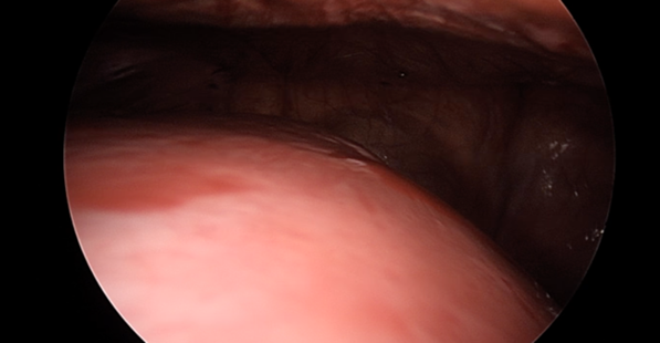
Figure 1: Intraoperative picture of the fibroid after supraumbilical access.
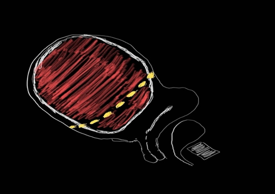
Figure 2: Sketch of the circumferential uterotomy.
The short way to the base of the fibroid is then reached shortly after, and the fibroid was completely removed with a part of the attached capsule in 34 min without opening the uterine cavity.
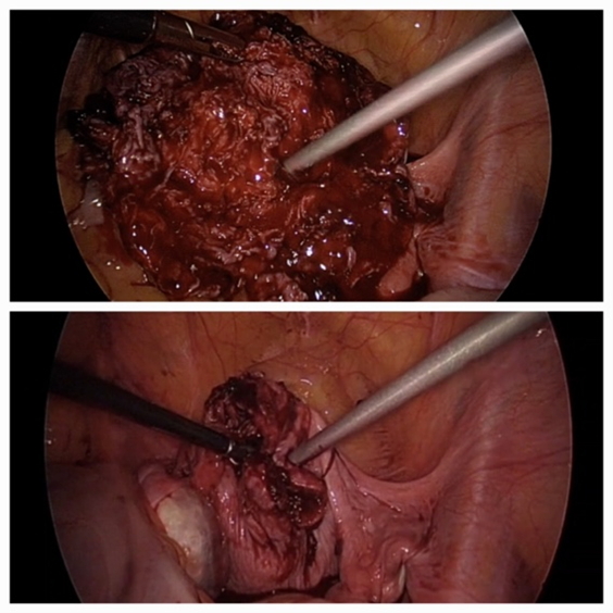
Figure 3: Intraoperative picture of the uterine posterior wall after removal of the fibroid.
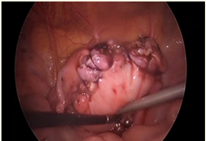
Figure 4: Intraoperative picture of the uterus after a multilayer reconstruction of the posterior wall.
A three-layer closure of the uterine incision was successfully performed to reconstruct the circular defect of the posterior uterine wall, and the fibroid was then morcellated in more than an hour.
The estimated Blood loss was approximately 180 ml intraoperatively. The postoperative weight of the morcellated fibroid was 1460 g.
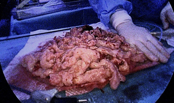
Figure 5: Picture of the fibroid after morcellation: 1460 g.
The postoperative recovery was fast. Two months after the operation, the sonographic scan showed a very good reformation of a full thickness posterior uterine wall.
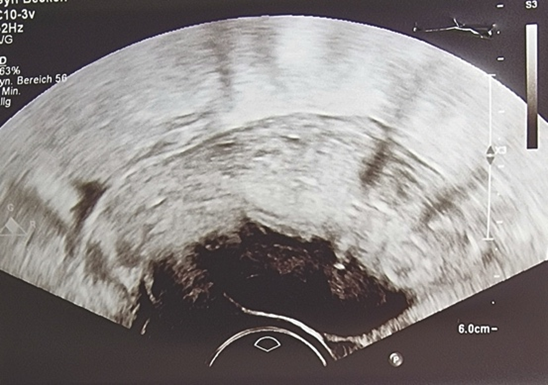
Figure 6: Ultrasound picture two months postoperatively showing a full thickness posterior uterine wall.
Laparoscopic myomectomy has gained popularity due to its minimally invasive nature, faster recovery, and reduced postoperative complications compared to traditional open surgery. However, its application for large fibroids (typically >10 cm in diameter) presents significant challenges, including limited visualization, increased technical difficulty, and longer operative times. Despite these obstacles, recent studies demonstrate that laparoscopic myomectomy can be successfully performed for large fibroids with the use of advanced techniques such as tissue morcellation, uterine manipulators, and vasopressin to reduce intraoperative bleeding [1]. The procedure allows for uterine preservation, making it an attractive option for women desiring future fertility [2].
Outcomes are generally favorable, with significant reductions in symptoms such as menorrhagia and pelvic pain, though the risk of complications, such as hemorrhage, remains higher compared to smaller fibroids [3]. Moreover, the surgeon's expertise plays a crucial role in reducing complications and achieving optimal results [4]. Studies show that, with proper patient selection and skilled surgical execution, laparoscopic myomectomy is a safe and effective option for large fibroids, offering the benefits of minimally invasive surgery along with acceptable complication rates and favorable reproductive outcomes [5].
There is no conflict of interest.
Copyright: © 2024 Mohamed Abuzaid. This is an open-access article distributed under the terms of the Creative Commons Attribution License, which permits unrestricted use, distribution, and reproduction in any medium, provided the original author and source are credited.