Mohamed Abuzaid*
Department of Gynaecology and Obstetrics, St. Vinzenz Hospital Hanau, Germany
*Corresponding Author: Mohamed Abuzaid, Department of Gynaecology and Obstetrics, St. Vinzenz Hospital Hanau, Germany.
Received: September 11, 2024; Published: September 24, 2024
Citation: Mohamed Abuzaid. “Laparoscopic Removal of Three Pelvic Splenosis Nodules Infiltrating the Right Pararectal Space: A Case Report". Acta Scientific Women's Health 6.10 (2024): 31-32.
Intraabdominal splenosis is a fascinating yet rare consequence of splenic rupture or splenectomy. While it is usually asymptomatic and discovered incidentally. In the rare cases where splenosis cause symptoms, surgical intervention may be required. Overall, the condition is benign, with a favorable prognosis for those affected.
Keywords: Laparoscopy; Spleen; Splenosis; Trauma; Splenectomy
The spleen is an essential organ in the human body, playing a critical role in filtering blood and managing the immune system. However, it is also susceptible to injury, particularly from blunt abdominal trauma, which can lead to a splenic rupture. In some cases, following a splenic rupture and subsequent splenectomy, an uncommon but notable condition can develop intraabdominal splenosis.
Splenosis represents the heterotopic autotransplantation of splenic tissue that usually follows traumatic rupture of the splenic capsule. More rarely, splenosis occurs after an iatrogenic seeding of splenic tissue during splenectomy. The splenic remnants implant on the serosal surfaces of the abdomen and chest, derive their blood supply from surrounding tissue [3].
During the traumatic event or surgery, fragments of the spleen can disseminate within the peritoneal cavity. These splenic fragments can then implant onto various surfaces, such as the omentum, mesentery, or other abdominal organs. Over time, these splenic tissues can vascularize and grow, forming splenosis. The process is mimicking tissue grafting, where the splenic cells adapt to their new location and continue to function [1].
26 years old Patient were referred for surgical removal of an adnexal Tumor on the right side. The tumor was discovered in the gynaecological examination due to the occasional lower abdominal pain of which the patient was complaining since few years. The Patient had a car accident 8 years ago with an urgent removal of a ruptured spleen.
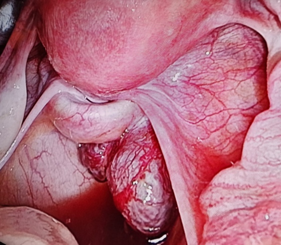
Figure 1: Intraoperative photo of three splenunculi in the pouch of douglas infilterating the right pararectal space.
Intraoperatively there were five fleshy tumors in the abdominal cavity. Three of them are in douglas pouch infiltrating the right pararectal space, one on the small intestine and the last one was impacted in the omentum. The whole five tumors were laparoscopicaly removed [4-6].
The histopathological examination resulted in five splenunculi in the three locations without malignancy.
There is no conflict of interest.
Copyright: © 2024 Mohamed Abuzaid. This is an open-access article distributed under the terms of the Creative Commons Attribution License, which permits unrestricted use, distribution, and reproduction in any medium, provided the original author and source are credited.
 Open Access by
Acta Scientific is licensed under a Creative Commons Attribution 4.0 International License
Open Access by
Acta Scientific is licensed under a Creative Commons Attribution 4.0 International License
Based on a work at https://actascientific.com
ff


