Dhurba DC1* and Ranjana Ranabhat2
1Veterinary Hospital and Livestock Service Expert Center, Hetauda, Makawanpur, Nepal
2United Veterinary Center, Hetauda, Makwanpur, Nepal
*Corresponding Author: Dhurba DC, Veterinary Hospital and Livestock Service Expert Center, Hetauda, Makawanpur, Nepal.
Received: May 12, 2021; Published: May 24, 2021
A four month old male buffalo calf with the history of bulge type swelling found at the point of umbilicus was brought to United Veterinary Center, Hetauda, Makawanpur, Nepal. Clinical examination revealed a reducible type small bulge mass with 4 fingers length and 3 fingers breadth hernial ring. Other clinical parameters were within the normal physiological limit. The case was corrected surgically by means of herniorrhaphy.
Keywords: Buffalo Calf; Bulge Type Swelling; Umbilicus; Herniorrhaphy
Hernia is the protrusion of an organ or tissue outside the abdominal cavity through natural or an abnormal opening [1]. A discontinuation of the abdominal wall at the region of umbilicus resulting protrusion of the abdominal contents in to hernia sac is known as umbilical hernia [5]. Some hernias are reducible, means hernial mass can be returned to their normal location through hernia ring while non-reducible type of hernia cannot pushed back [1]. Hernia may be congenital or acquired [7]. In calf, umbilical hernia is commonly present due to failure of the normal closure of umbilical ring [4]. From such incomplete closure of umbilical ring the abdominal contents like intestine, omentum comes out [3]. Acquired cause of umbilical hernia includes weak abdominal wall (imperfect occlusion of umbilicus), deep wound, abscess, increased intra-abdominal pressure due to straining due to constipation or diarrhea, violent exercise etc [2].
This condition can be diagnosed by external digital palpation, clinical observation, or ultrasonography [4]. There are different methods for the correction of umbilical hernia such as ligation of hernia sac, suturing of hernia ring, use of clamps and surgical correction [8]. But if the hernia is greater than 5 cm then the surgical correction (herniorrhaphy) is the treatment of choice for the correction of umbilical hernia [6].
A male buffalo calf of 4 months was brought to United Veterinary Center, Hetauda with the history of bulge type swelling found at the point of umbilicus. According to owner bulge mass was present since birth and which tends to increase in size with age. The palpation at the site of swelling confirmed that the herinal mass is reducible type. The size of hernial ring was 4 fingers length and 3 fingers breadth. Clinical parameters such as heart rate, respiratory rate and temperature were within normal physiological range. No any other behavioral changes were noticed.
Surgical correctionThe calf was restrained on right lateral recumbence. Cranial paravertebral regional nerve block was done by using 2% lignocaine. Then, the site of incision (umbilical area) was prepared for aseptic surgery by shaving, scrubbing, washing with Salvon® (Cetrimide 3% and Chlorhexidine gluconate 1.5% solution). At operative site 2% lignocaine was infiltrated locally. After proper analgesia the animal was controlled in dorsal recumbency and about 7 cm long longitudinal midline incision was made over swelling mass. The skin was detached from subcutaneous tissues and the incision was continued through abdominal muscles and peritoneum avoiding blood vessels.
A part of small intestine was found as hernial content. Gently the intestine was repositioned into abdominal cavity through the umbilical ring. The hernial ring was closed by overlapping mattress suture after freshened the edges of hernial ring. Absorbable suture material (No. 2 catgut) was used to close the opening. The subcutaneous layer was sutured with No 2 catgut and skin with Nylon in interrupted suture pattern.
Recommended post-operative care Systemic antibiotic:Regular dressing with 5% povidone iodine for 10 days.
Finally, on 12 day post-operative complete healing was recorded and the skin suture were removed on the same day.

Figure 1: Swelling at the point of umbilicus.
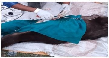
Figure 2: Midline longitudinal incision at the site hernia.

Figure 3: Interrupted skin suturing.

Figure 4: Recovered calf.
In a male buffalo calf the umbilical hernia was corrected successfully with herniorrhaphy. After proper diagnosis, timely operation and good post-operative care is major for the correction of umbilical hernia.
The author declare that there is no any conflict of interest.
Citation: Dhurba DC and Ranjana Ranabhat. “Surgical Correction of Umbilical Hernia in Buffalo Calf: A Case Report”.Acta Scientific Veterinary Sciences 3.6 (2021): 33-35.
Copyright: © 2021 Dhurba DC and Ranjana Ranabhat. This is an open-access article distributed under the terms of the Creative Commons Attribution License, which permits unrestricted use, distribution, and reproduction in any medium, provided the original author and source are credited.


 Open Access by
Acta Scientific is licensed under a Creative Commons Attribution 4.0 International License
Open Access by
Acta Scientific is licensed under a Creative Commons Attribution 4.0 International License
Based on a work at https://actascientific.com
ff
