Aman1, Vikasdeep Gupta2*, Ashiya Goel3, Nikhil Rajan2 and Ankur Mohan2
1Department of Otolartyngology & Head Neck Surgery, Dr Aman ENT and Cancer Clinic Jhajjar, India
2Department of Otolartyngology & Head Neck Surgery, All India Institute of Medical Sciences, Bathinda, India
3Department of Otolartyngology & Head Neck Surgery, Pragma Medical Institute, Bathinda, India
*Corresponding Author: Vikasdeep Gupta, Associate Professor, All India Institute of Medical Sciences (AIIMS), Bathinda (Punjab), India
Received: January 06, 2024; Published: September 27, 2024
Citation: Vikasdeep Gupta., et al. “Pleomorphic Adenoma of Anterolateral Tongue: A Rare Case Report". Acta Scientific Otolaryngology 6.10 (2024):36-38.
Pleomorphic adenoma is the commonest benign tumor of salivary glands [1-3]. The parotid gland being most frequent to be involved site followed by submandibular in major salivary glands, whereas palate in minor salivary glands followed by lips. A 41 yr old male presented to the ENT OPD with swelling on the left side of the anterior aspect of the tongue. Pleomorphic adenoma of tongue is a very rare tumor of the tongue with high chances of recurrence.
Keywords: Pleomorphic Adenoma; Salivary Glands; Tongue
Pleomorphic adenoma is the commonest benign tumor of salivary glands [1-3]. The parotid gland being most frequent to be involved site followed by submandibular in major salivary glands, whereas palate in minor salivary glands followed by lips [4,5]. Tongue is an infrequent site for its occurrence. Out of the 26 cases reported till now, the posterior part of the tongue with 19 cases showed the highest incidence followed by anterior with 5 cases and lateral with 2 cases, respectively. It presents as a swelling over the tongue, dysphagia, foreign body sensation in the mouth, and difficulty articulating words. Its first line treatment is wide surgical excision with 1 cm margins. In this report, we are presenting a case of pleomorphic adenoma of tongue in a 38-year-old male in which wedge shaped wide surgical excision was done.
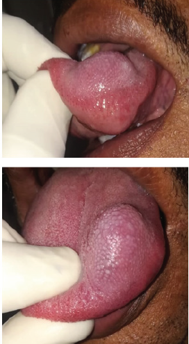
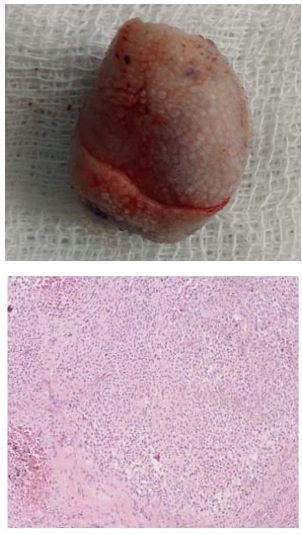
A 41 yr old male presented to the ENT OPD with swelling on the left side of the anterior aspect of the tongue for 12 months which was insidious in onset and gradually progressive. No complaints of pain, bleeding, or difficulty in swallowing. No history of trauma to the tongue. On local examination, there was a smooth surface swelling of approximate size 4*2 cm on the left side of anterior aspect of the tongue. Overlying mucosa was normal. On palpation, the swelling was firm and non-tender. Rest of the oral cavity and oropharynx was unremarkable. Indirect laryngoscopy was normal. No cervical lymphadenopathy was palpated. Fine needle aspiration cytology revealed cytological features suggestive of salivary gland neoplasm with closest resemblance with pleomorphic adenoma. The patient was planned for transoral wedge-shaped surgical excision of the tumor under general anesthesia. The patient underwent the same with no immediate postoperative complications. On histopathological examination, section revealed stratified squamous epithelium covered soft tissue revealing a well circumscribed tumor comprising polygonal cells with large chondromyxoid areas. On immunohistochemistry, tumour cells are GFAP and vimentin-positive, CK negative and CD31 negative and positive in vascular endothelial cells, possibility of a salivary gland neoplasm, closest resemblance to pleomorphic adenoma.

Figure 1: Showing mass on the anterior tongue.

Figure 2: Showing excised on gross and HP examination.
The tongue has a propensity for occurrence of malignant tumors more than benign ones with a ratio of 1:6 [2]. Pleomorphic adenoma is the most common benign tumor. It is seen most commonly in the posterior part of the tongue, followed by anterior and lateral. Since 1960, only 26 cases of pleomorphic adenoma of the tongue have been reported. Out of which, 5 cases were from the anterior tongue, 2 cases from the lateral side, and 19 cases from the posterior tongue. Histologically, it is a mixed tumor because it contains both epithelial and myoepithelial components. It is a slow-growing painless tumor with a relatively late presentation. It can occur between 9-90 years old, but the 4th to 5th decade is the most common age of presentation. Females are affected more commonly than males [4]. It is of 3 histological subtypes, myxoid (80% stroma), cellular with more myoepithelial cells and mixed. It is an encapsulated tumour [6-8].
Treatment of choice for it is wide surgical excision with approximately 5 mm margins and long follow up because of high risk of its late recurrence [9]. Recurrence may be due to tumour spillage, inadequate surgical excision, or capsular rupture during surgery. Incomplete capsules with tumour cells next to mucosa may also be seen in intraoral pleomorphic adenoma, which demands removal of overlying mucosa for excision of tumour to reduce recurrence [10]. To prevent tumour spillage, cutting into the tumour should be avoided. In case of tumor spillage, proper washing of the wound with the removal of as much as possible tissue with postoperative radiotherapy must be given [11-15]. It can convert into carcinoma ex pleomorphic adenoma if left untreated.
Pleomorphic adenoma of tongue is a very rare tumor of the tongue with high chances of recurrence. Wide surgical excision with around 5 mm to 1cm margins is the treatment of choice. While considering differential diagnosis, carcinoma ex pleomorphic adenoma must be kept in mind.
No conflict of interest is there.
Copyright: © 2024 Vikasdeep Gupta., et al. This is an open-access article distributed under the terms of the Creative Commons Attribution License, which permits unrestricted use, distribution, and reproduction in any medium, provided the original author and source are credited.