Buzdugan IO*, Belkovskyi IA, Petrashchuk TB, Puiu VI, Slyvchuk DV, Stankevych IA, Tsurkan PE, Chornyi OV, Shurhaliuk KS, Bukach OP, Voloshuna LO, Ratsa VV and Zolotyn IM
Department of internal Medicine, Bukoviynian State Medical University, Ukranie
*Corresponding Author: Buzdugan IO, Department of internal Medicine, Bukoviynian State Medical University, Ukranie.
Received: August 20, 2024; Published: September 19, 2024
Citation: Buzdugan IO., et al. “Assessment of Dyspeptic and Pain Syndromes Depending on the Size of the Peptic Ulcer of the Stomach". Acta Scientific Gastrointestinal Disorders 7.10 (2024):16-19.
Introduction: The article presents clinical features of dyspeptic and pain syndromes of peptic ulcer of the stomach. These pathology data remain an unsolved question and are relevant today.
The purpose of the study is to assess dyspeptic and pain syndromes depending on the size of the peptic ulcer.
Research Materials: All patients received inpatient treatment in the gastroenterology department of the Chernivtsi Regional Clinical Hospital in 2013-2016. The diagnosis was carried out in accordance with the National recommendations of the Unified Clinical Protocol of primary, emergency and secondary (specialized) medical care for patients with PVS and DPK in adults. approved by order of the Ministry of Health of Ukraine No. 613 of the Ministry of Health of Ukraine dated September 3, 2014.
Research Results: The analysis of the features of the clinical course of the disease showed that all patients infected with HP had a pain syndrome, which was noted by 100% of patients with PU. The most frequent localization of pain was in the epigastric region (64.7%) of a dull aching nature (58.8%) with a decrease in pain after eating (70.6%). It was established that the average degree of severity (CCT) of the pain syndrome in patients with PU with the presence of CagA+VacA+ strains was 2.18, and in the combination CagA+VacA-/CagA-VacA+ ─ 2.
The dyspeptic syndrome was accompanied by loss of appetite (70.6%), nausea (70.6%) with the spread of emotional lability (70.6%) (р<0.05). The highest CCT of dyspeptic syndrome is found in patients with CagA+VacA+ PVS (2.35 points) with a disease duration of up to 5 years in 57.58% of the examined persons, which characterizes the severity of the course and increased pain syndrome in the presence of both toxigenic strains.
Conclusion: Therefore, the increase in pain and astheno-vegetative syndromes with a decrease in dyspepsia depends on the erosive defect of the mucosa caused by the combination of toxigenic strains of CagA+VacA+ Helicobacter infection in patients with peptic ulcer.
Keywords: Peptic Ulcer; Stomach; Pain; Dyspepsia; Syndrome
As you know, peptic ulcer (PU) is one of the most common diseases in modern gastroenterology [1,3]. And the influence of H. Pylori, as the main pathogen, has been proven long ago and convincingly. However, over time, there have been reports about the immune defense of the host organism, which, together with the factors of the stomach environment, create a high rate of genomic variability of H. pylori [2,5], which contributes to the active evolution of the pathogen and its adaptation to its host. The increased variability of HR (genes сagA and vacA) leads to the development of the most serious diseases of the stomach and gastrointestinal tract: atrophic gastritis, peptic ulcer disease, and stomach cancer [4,6]. The purpose of the study is to assess dyspeptic and pain syndromes depending on the size of the peptic ulcer.
All patients received inpatient treatment in the gastroenterology department of Chernivtsi Regional Clinical Hospital in 2013-2016.
The presence of PVS was verified on the basis of the clinical picture, anamnesis data, objective methods of examining the patient, the results of endoscopic and morphological studies of OSH.
On the basis of patients' complaints and the results of an objective examination, we estimated the indicator of average degree of severity (CCT). For this, we used data on the intensity of pain on a scale where 0 points mean the absence of complaints; 1 point - minimal complaints; 2 points- moderate complaints; 3 points — pronounced or very pronounced complaints. The SST was estimated according to the formula:
where CCT is the average degree of severity of clinical manifestations; a - the number of patients with symptom severity of 1 point; b - the number of patients with symptom severity of 2 points; c - the number of patients with symptom severity of 3 points; d - the number of patients with no symptom.
Diagnostics was carried out in accordance with the National recommendations of the Unified clinical protocol of primary, emergency and secondary (specialized) medical care for patients with PU in adults, approved by the order of the Ministry of Health of Ukraine No. 613 of the Ministry of Health of Ukraine dated September 3, 2014.
The study did not include patients under the age of 18 and older than 75 years with HP-negative and complicated (perforation, penetration, bleeding, etc.) PU of the stomach.
Figure 1 shows the distribution of peptic ulcer of the stomach depending on localization.
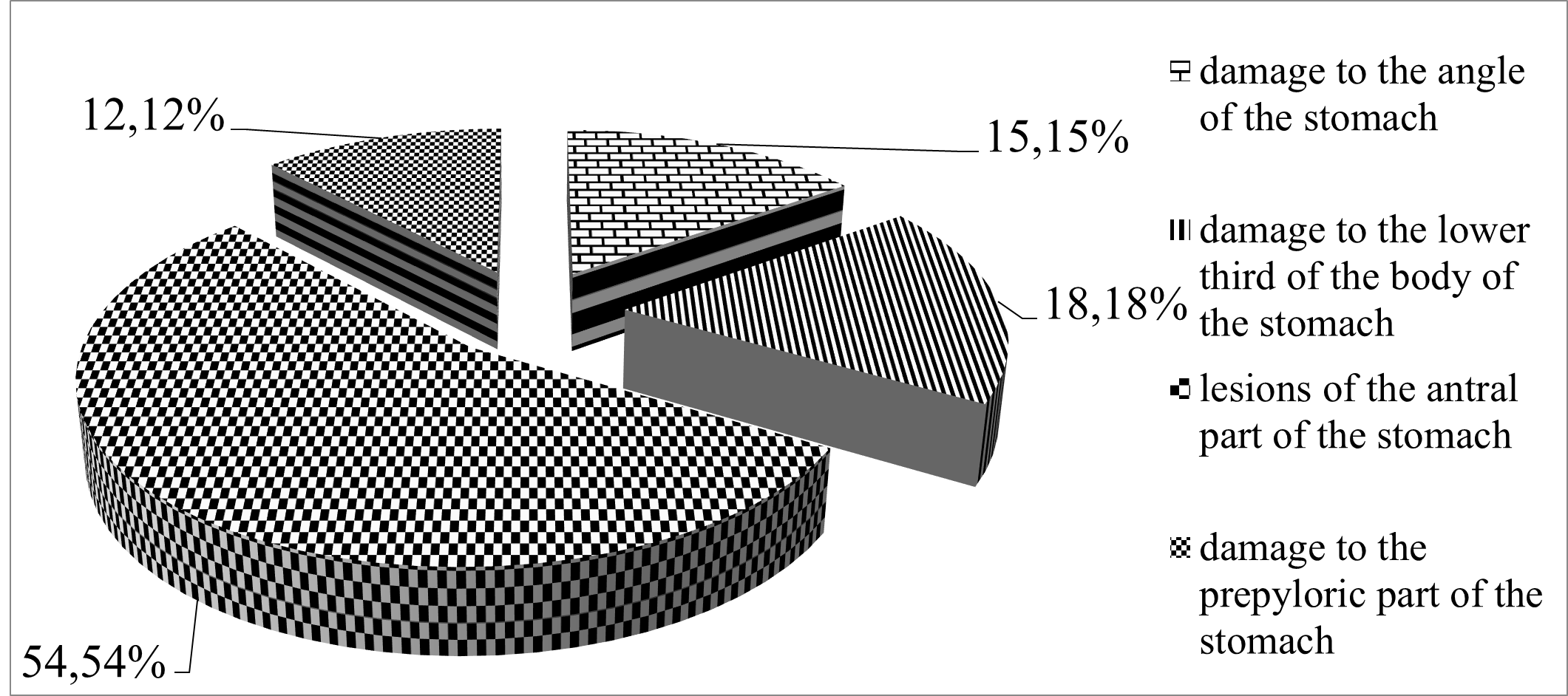
Figure 1: Distribution of patients with peptic ulcer of the stomach depending on localization of the ulcer defect, %.
In patients with PVS, a small-sized ulcer was observed in 54.5% of patients, and medium and large-sized ulcers were observed in 39.4% and 6.1% of the studied persons (Table 1).
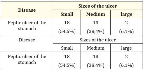
Table 1: Distribution of patients with peptic ulcer of the stomach depending on the size of the ulcer, (number of people).
Evaluating the duration of the disease (Table 2), it was established that in patients with PU, frequent detection of ulcers with a duration of up to 5 years is observed in 19 patients (57.58%) and first detected - in 7 patients (21.21%).
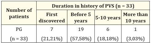
Table 2: Distribution of patients with PVS according to the duration of the disease.
The analysis of the features of the clinical course of the disease (Table 3) showed that all patients infected with HP had a pain syndrome. The presence of pain was noted among patients with PVS. The most frequent localization of pain was in the epigastric region (64.7%) of a dull aching nature (58.8%) with a decrease in pain after eating (70.6%).
The dyspeptic syndrome was accompanied by loss of appetite (70.6%), nausea (70.6%) with the spread of emotional lability (70.6%) (р < 0.05).
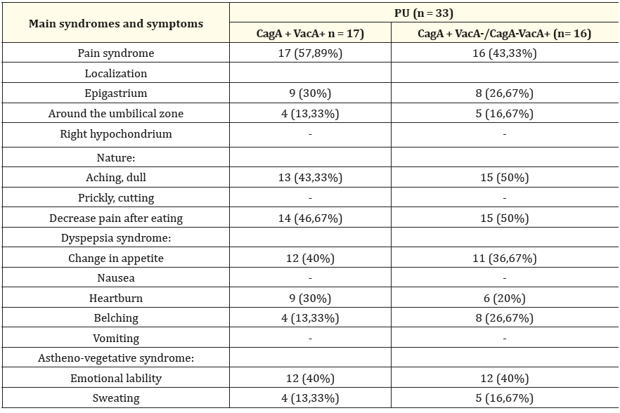
Table 3: Clinical characteristics of peptic ulcer of the stomach (number of patients, %).
Evaluating the clinical picture of PU, it was established that the pain syndrome was detected in 100% of patients. However, the intensity of pain and its average degree of severity also depends on the presence of a combination of toxigenic strains of H. pylori (Figure 2).
It was established that the average degree of severity (CCT) of the pain syndrome in patients with PVS with the presence of CagA + VacA+ strains was 2.18, and in the combination CagA + VacA-/CagA-VacA+- 2.
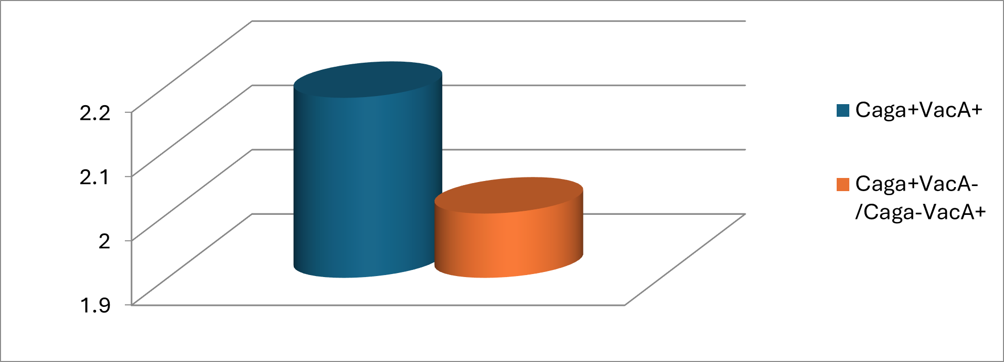
Figure 2: Intensity of pain syndrome in patients with PU taking into account the toxigenicity of H. pylori strains.
The presence of dyspeptic syndrome is a characteristic feature of peptic ulcer. However, in the presence of PVS, taking into account the toxigenicity of H. pylori strains of CST, the degree of dyspeptic syndrome is different (Figure 3).
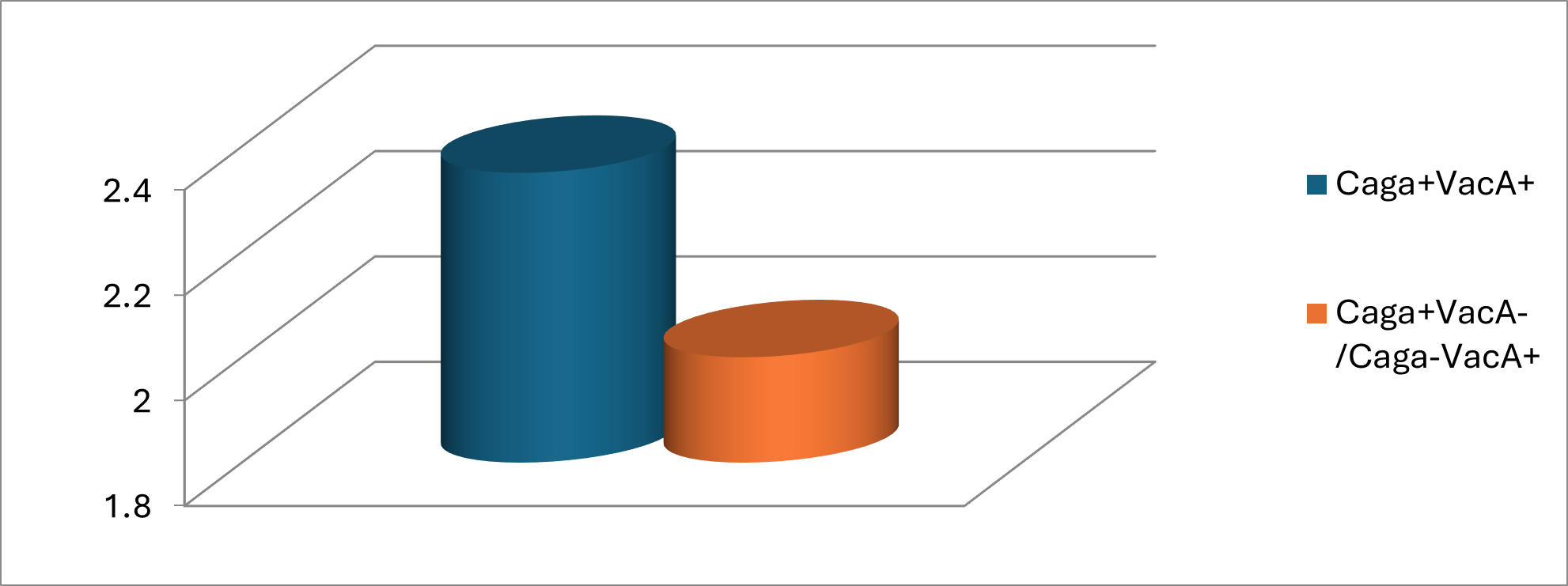
Figure 3: Intensity of dyspeptic syndrome in patients with PU taking into account the toxigenicity of H. pylori strains.
The highest CST of dyspeptic syndrome occurs in patients with CagA + VacA+ PU (2.35).
No less important is the presence of astheno-vegetative syndrome, manifested by emotional lability and sweating. However, the presence of toxigenic strains, their combinations changed the manifestation of astheno-vegetative syndrome and burdened CCT.
In patients with the presence of CagA + VacA-/CagA-VacA+ strains, manifestations of the astheno-vegetative syndrome are less pronounced compared to the group of patients with the presence of both toxigenic CagA + VacA+ strains.
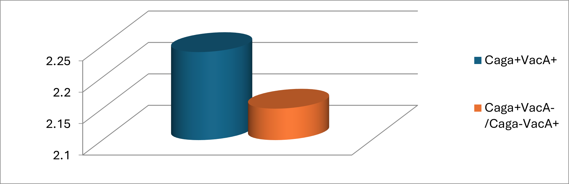
Figure 4: Intensity of astheno-vegetative syndrome in patients with PU taking into account the toxigenicity of H. pylori strains.
Therefore, the increase in pain and astheno-vegetative syndromes with a decrease in dyspepsia depends on the erosive defect of the mucosa caused by the combination of toxigenic strains of CagA + VacA+ Helicobacter infection in patients with peptic ulcer.
Copyright: © 2024 Buzdugan IO.,et al This is an open-access article distributed under the terms of the Creative Commons Attribution License, which permits unrestricted use, distribution, and reproduction in any medium, provided the original author and source are credited.
 Open Access by
Acta Scientific is licensed under a Creative Commons Attribution 4.0 International License
Open Access by
Acta Scientific is licensed under a Creative Commons Attribution 4.0 International License
Based on a work at https://actascientific.com
ff


