Nitu Sharma*
Radiology, Jr Consultant, Head of Department, BP Koirala Memorial Cancer Hospital, Nepal
*Corresponding Author: Nitu Sharma, Radiology, Jr Consultant, Head of Department, BP Koirala Memorial Cancer Hospital, Nepal.
Received: August 20, 2024; Published: September 05, 2024
Citation: Nitu Sharma. “Comparative Evaluation of the Bowel Wall Appearance and Bowel Distention by Using Water Contrast and Commercially Prepared Agent (Iopamidol) of the Upper Gastrointestinal Tract in Multiaxial CT scans: BPKMCH Experience". Acta Scientific Gastrointestinal Disorders 7.10 (2024):03-07.
Objective: This study aims to evaluate the performance of an orally administered water contrast versus commercially prepared (Iopamidol) contrast in the upper gastrointestinal tract CT scan. To be able to improve the diagnosis of abdominal abnormalities in multi axial CT scans.
Materials and Methods: Sixty patients were seen during the twelve-month research study. Thirty patient used water contrast and 30 patient used commercially prepared (Iopamidol) contrast in the abdominal CT scan. The first group was given a volume of one litre of water as contrast one hour before the procedure and additional two hundred to two hundred fifty (200-250 cc) water was given fifteen to thirty minutes before the start of the procedures. The second group was given a solution containing thirty cc of Iopamidol mixed to one litre of water one litre given one hour before the procedure, and additional two hundred to two hundred fifty cc, fifteen to thirty minutes before the start of the procedure. The degree of distention and the visualization of the mural detail was qualitatively scored on five-point scale. The differences were evaluated by using Mann-Whitney test.
Results: There were no statistical difference in the distention and visualization of the stomach , duodenum, jejunum, and ileum (P = 0.5671) in group 1 and group 2. In addition, there are no significant difference in the distention comparing the cross-sectional diameter in duodenum (P value = 0.351), jejunum (P value = 0.305), and ileum (P = 0.5569).
Conclusion: Water can also be an effective contrast medium for distension and excellent visualization of mural features in the gastrointestinal tract. The use of water as oral contrast is universally available and cost effective compared to the commercially prepared (Iopamidol) contrast.
Keywords: Bowel Wall; Bowel Distention; Iopamidol; Gastrointestinal Tract; BPKMCH
Computerized tomography, and often formerly referred to as computerized axial tomography (CAT) scan, is an X-ray procedure that combines many X-ray images with the aid of a computer to generate cross-sectional views and, if needed, three-dimensional images of the internal organs and structures of the body [1]. The small bowel has always been a challenging area to assess for surgeons and gastroenterologists owing to its long length and complexity of the loops. Technological advances in multi-detector computed tomography (MDCT) have revolutionized imaging field and have added new concepts to solid and hollow viscera imaging [2]. The success of accurate interpretation of bowel pathologies requires an optimal preparation and acquisition. Luminal distension and fold visualization are the determining factors in gastrointestinal tract imaging. This requires an oral contrast agent, which should cause uniform intraluminal attenuation, high contrast between luminal content and bowel wall, absence of artifact formation and no significant adverse effects [3]. Neutral endoluminal contrast could potentially improved diagnosis of abdominal abnormalities at multi axial CT scans. Nuetral contrast have been shown to be valuable in the diagnosis of small bowel disorders and have been used to marked the stomach and duodenum during evaluation of pancreas and biliary tree [4]. Water is an excellent contrast agent when used during upper abdominal CT scanning, but because water is rapidly absorbed throught the intestinal wall, the used of contrast agent in jejunum and ieum is limited. We will compare the performance of two contrast agents: water and commercially prepared contrast (Iopamidol) to improve bowel distension.
The advantage of water as oral contrast is its easy availability and affordability compared to the commercially prepared oral contrast (Iopamidol).
Prospective study
Descriptive study
The study was done in BPKMCH, a tertiary hospital with 450 bed capacity located in Bharatpur, Chitwan Nepal.
All BPKMCH’S admitted and outpatient who underwent Abdominal CT scan from January 2022 to December 2023.
This study was a prospective, descriptive study. The sample size was determined by the number of patient who underwent abdominal CT scan which were able to comply with the set guidelines in inclusion criteria.
Image will be obtained using fujifilm 128 slices.
Each subjects were assigned with personal case number. Two attending radiologists with experience in body imaging will independently review the 60 patients included in this study. Readers rated each image on its clarity of anatomical detail on segments of the gastrointestinal tract (gastric fundus, gastric antrum, duodenal folds, jejunal and ileal folds) in five-point scale (0 = worst, 4 = best).
From January to December 2023, 60 patients (30 men and 30 women; age range 20-40 years; mean age of 30 years old was scheduled for abdominal CT in whom oral contrast (water) and commercially prepared contrast (Iopamidol) was given. All patient enrolled in this study gave informed consent.
A total of 60 inpatients and outpatients (30 women, 30 men; average age of 30 years) who were known to have or were suspected of having pancreatic or biliary disease were referred for abdominal CT scan. Patient was randomized into two groups on the basis of the type of water oral contrast agent versus commercially prepared (iopamidol) contrast study. Group 1 received 1 liter or water contrast agent one hour before the procedure, and additional 200-250 cc of water was given 15-30 minutes before the procedures. The group 2 was given a solution containing agent (30 cc of iopamidol mixed to 1L of water), 1liter was given 1 hour before the procedure and additional 200-250 cc, 15-30 minutes before the procedure.
The studies was reviewed by two certified radiologist. Because of the high attenuation of the contrast (Iopamindol) being compared to neutral (water) intraluminal contrast, it was impossible to blind the oral contrast administered. They graded the gastrointestinal tract distention and mural visualization of the scans obtained in the stomach, duodenum, jejunum, and ileum. They used five-point scale (0 = none, 1 = poor, 2=partial, 3 = good, 4-5 = full distention and mural visualization.
Specific indications for this study was patient who are suspected or known case of pancreatic disease. Age group of 20-40 years old with mean age of 30 years old. Complaints of abdominal pain (epigastric area). Patient was excluded in the study if known to have undergone prior surgery.
In order to determine the statistical significance for qualitative analysis, an arithmetic mean were recorded by each group for luminal distention and visualization of the stomach, duodenum, jejunum and ileum was calculated. The arithmetic mean value for each group was assessed by visual distention in the stomach, duodenum, jejunum and ileum. Visualization was judge based on the reader’s ability to delineate the mural anatomic features that were appropriate for each specific segment. The gastric visualization was based on the scores for the uniform thickness in the wall of the gastic fundus and antrum. Visualization of the ileum was to delineate the ileal wall. The qualitative data for distention and visualization in each segment for each patients were averaged for 2 groups.
For the quantitative analysis a mixed-model squares regression was used to examine the differences of the contrast agent observed in each patient in each three region of the small intestines (duodenum, jejunum, and ileum).
Abdominal CT scan was able to demonstrate 25 abnormalities out of 60 patients (41%) (Table 1). There were 11 pancreas related abnormalities, including seven pseudocyst, 3 acute pancreatitis and 1 chronic pancreatitis. There were concurrence between the two readers in 10 patient. In one patients, reader 2 interpreted the findings as chronic pancreatitis. Abnormalities that were not related to pancreas were observed in 14 patients, including 8 lymphomas, 6 liver masses. The resulting p value of 0.728 denotes that there exist no significant difference between the two groups in terms of abnormalities related and non-related to pancreas.Normal Abdominal CT scan were noted in the 35 patient (58%).
For age and sex (Table 2) for distention of bowel segments a total of 30 women and 30 men (mean age of 30) receives water contrast agent versus commercially prepared (Iopamidol) contrast agent. There were no statistically significant difference between those who received water and commercially prepared (Iopamidol).
For qualitative analysis (Table 3) the mean values for distension in the stomach (P = 1.000) , duodenum (P = 0.35ly 1), jejunum (P = 305), ileum (P = 0.5569) were significantly the same in group 1 and 2. The mean score for the visualization of anatomic detail in duodenum (P = 0.351), jejunum (P = 0.305) , ileum (P = 0.5569) were significantly the same in both group 1 and group 2. The scores in group 1 is almost the same in group 2.
The result of quantitative analysis (Table IV) of distention in the duodenum, shows there were no significant relationship between contrast agent used and the age (P value = 0.351).
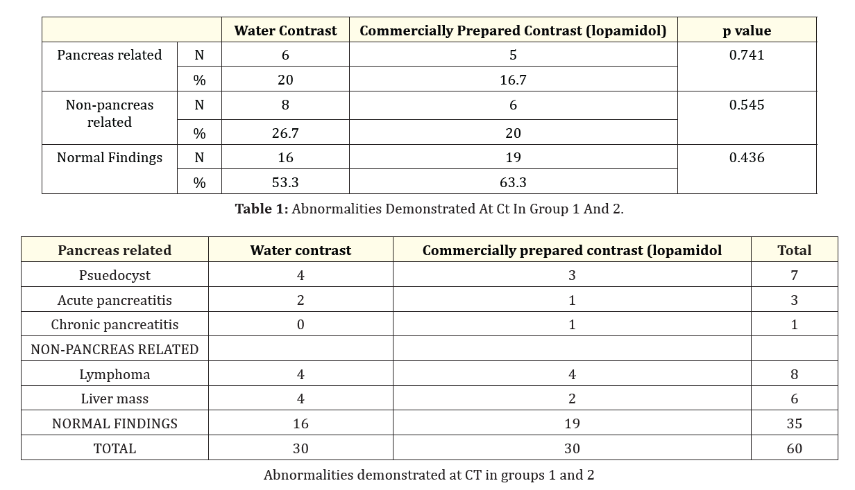
Table 1: Abnormalities Demonstrated At Ct In Group 1 And 2.
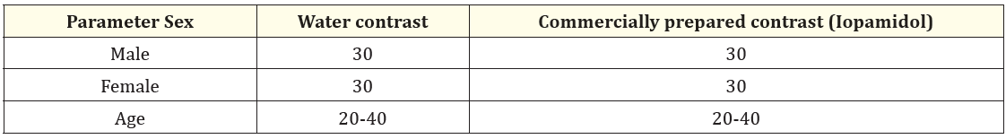
Table 2: Classification of patients according to age and sex.
P value = 1.000 for male and female
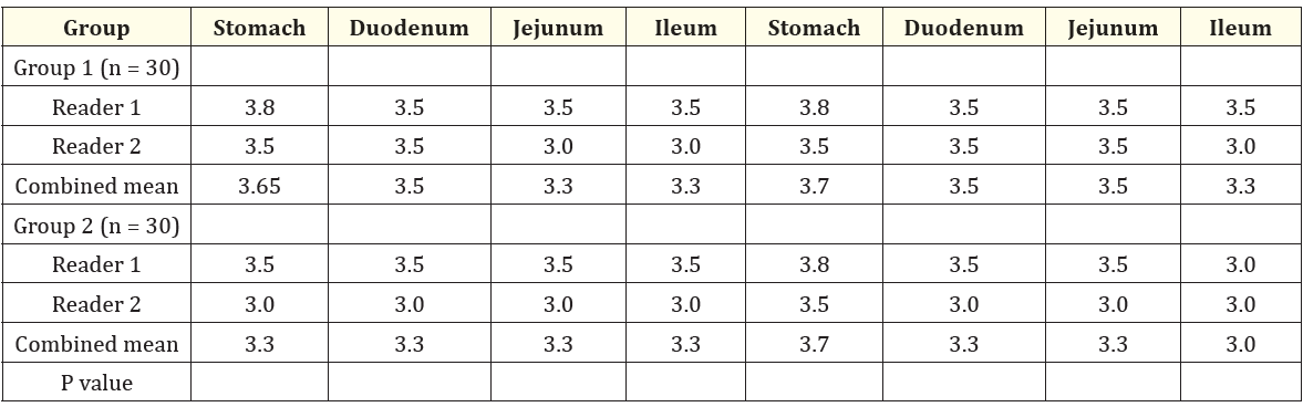
Table 3: Results of qualititative anaylis in small intestines for each group.
Distention Visualization
For qualitative analysis,distension and visualization were scored using a continous five point scale (0 = worst), 4 = best)
For group 1, 1 liter of water contrast was administered, and for group 2, The group 2 was given a solution containing agent
(30 cc ofiopamidol mixed to 1L of water.
The combined mean was calculated as the arithmetic mean of both readers.
P values were calculated using the Mann-whitney test.
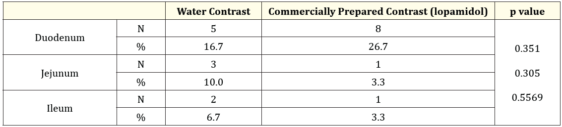
Table 4: Results of quantitative analysis for each contrast agent.
An increased interest in the used of neutral oral contrast agents has paralleled the widespread clinical use of multi– detector row CT scanners [5-8]. It should be noted, however, that the rapid drinking of the neutral contrast agent achieved a major goal that is, the reproducibly of uniform distention along the length of the gastrointestinal tract. The capacity to visualized the bowel wall and delineate the lumen adds the efficiency of abdominal CT scan examination. By using oral contrast agent and solution containing contrast agent enable the radiologist to routinely conceptualized the gastrointestinal tract.
Water as oral contrast agent compared to the commercially prepared (Iopamidol) contrast in abdominal CT-scan were able to delineate the stomach (P value = 1.000). Both contrast agent demonstrate significantly full distention in the segments of the gastrointestinal tract and show good anatomic detail in the duodenum, jejunum, and ileum. The tolerability and palatability of both contrast agents are comparable. All participants regardless of age and sex were able to cooperate and comply for the requirements of the procedure.
Using present CT scan technology, water can also be effective contrast medium causing better of equal distension and excellent visualization of mural features in the gastrointestinal tract. The water oral contrast is easily available and cost effective compared to the commercially prepared (Iopamidol) contrast. Using water as contrast agent for upper abdominal CT scan is therefore recommended especially for the evaluation of the upper abdominal CT scan.
Copyright: © 2024 Nitu Sharma. This is an open-access article distributed under the terms of the Creative Commons Attribution License, which permits unrestricted use, distribution, and reproduction in any medium, provided the original author and source are credited.
 Open Access by
Acta Scientific is licensed under a Creative Commons Attribution 4.0 International License
Open Access by
Acta Scientific is licensed under a Creative Commons Attribution 4.0 International License
Based on a work at https://actascientific.com
ff


