Vipul Garg1, Vanshikha Sharma1*, Dakshanya Chatterjee1, Ridam Bhasin2, Parveen Dahiya2, Megha Dhadwal1 and Shruti Thakur1
1Department of Oral and Maxillofacial Surgery, Himachal Institute of Dental Sciences, India
2Department of Periodontology and Implantology, Himachal Institute of Dental Sciences, India
*Corresponding Author: Vanshikha Sharma, Department of Oral and Maxillofacial Surgery, Himachal Institute of Dental Sciences, India.
Received: October 08, 2024; Published: October 31, 2024
Citation: Vanshikha Sharma., et al. “Beyond the Ordinary: Neurilemmoma Unveiled in The Mental Nerve". Acta Scientific Dental Sciences 8.11 (2024):79-82.
Neurilemmoma, alternatively referred to as schwannoma, is a benign tumour of the nerve sheath that seldom develops in the oral cavity. It appears as a encapsulated, slow-growing, single, smooth-surfaced tumour that typically does not cause symptoms. We report a case of 15-year-old male with a well-defined mass on left mandibular alveolar mucosa in the premolar region which was painless and slow growing. Diagnosis is made by clinical and histopathological examination to confirm the neural tissue origin of the lesion. The treatment is complete surgical excision of the lesion without recurrence.
Keywords: Schwannoma; Neurilemmoma; Mental Nerve; Mandible; Excision
Schwann cells form a thin outline around each extracranial nerve fiber and wrap larger fibers with an insulating membrane, myelin sheath, to enhance nerve conductance. Schwannomas arise when proliferating Schwann cells form a tumor mass encompassing motor and sensory peripheral nerves [1]. A neurilemmoma, or schwannoma, is a benign tumor of the nerve sheath that infrequently develops in the oral cavity. Only 1% of all schwannomas outside the skull occur in the mouth, with the tongue being the most frequent location and the retromolar region being the least common. These tumors appear as encapsulated, slow- growing, single masses with a smooth surface, typically causing no symptoms [2]. Therefore, schwannoma, known by several names including neurilemmoma, neurinoma, and perineural fibroblastoma, is a benign tumor that grows slowly and is encapsulated. It originates from Schwann cells of the neural sheath, affecting peripheral, cranial (excluding optic and olfactory), spinal, and autonomic nerves [1,3].
Upon histologic examination, the clearly delineated neurilemmoma exhibits degenerative alterations and varying patterns: dense spindle- shaped structures termed "Antoni A" areas, alongside microcystic regions containing numerous macrophages and collagen fibers known as "Antoni B" areas [4].
The presence and intensity of symptoms are determined by the size and location of the lesions. Typically, these swellings are asymptomatic, single, with a smooth surface, and exhibit slow growth. Periodically, they may cause tenderness or irritation. The primary goal of treatment is complete surgical removal, which greatly reduces the likelihood of recurrence [1].
A 15 year old boy presented to the Department of Oral and Maxillofacial Surgery, HIDS, Paonta Sahib on 13th June 2023 with the chief complaint of a swelling in the lower left front tooth region since 6 months. The patient was apparently alright 6 months ago, when he suddenly developed a painless slow growing nodule present at the junction of the attached gingiva and alveolar mucosa in the left mandibular premolar region. Clinical examination revealed a nodule of 3x3cm in size, cylindrical in shape, non tender, smooth in texture and rubbery on palpation. The overlying mucosa was slightly erythematous with no cervical lymphadenopathy. His general condition and nutritional status were good. The patient was otherwise healthy and had no significant past medical history and no other symptoms; his family history was also unremarkable.
Clinically the lesion appeared to be benign soft tissue neoplasm and provisional diagnosis of intraoral lipoma was suggested. The complete blood count gave results within normal limits.
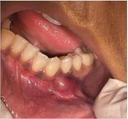
Figure 1: Asymptomatic nodule in the left mandibular mucosa.
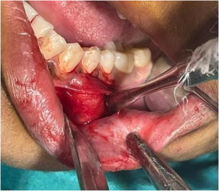
Figure 2: Incision placed and blunt dissection done.
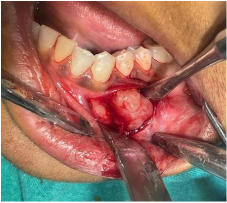
Figure 3: Excisional biopsy of the nodule.
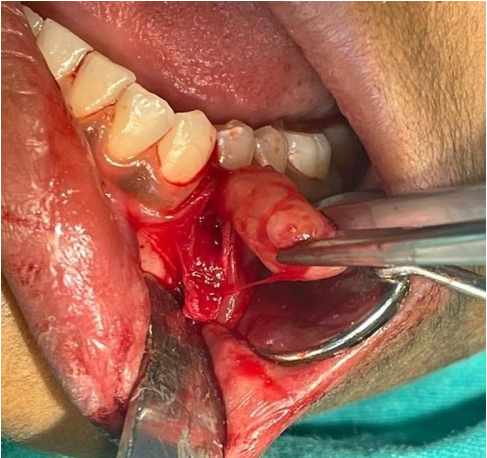
Figure 4: Preservation of the mental nerve.
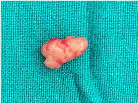
Figure 5: Excised Specimen.
Considering the size of the lesion an excisional biopsy was planned under local anesthesia. Vestibular incision was given along the length of the lesion from canine to second premolar. The lesion was then carefully excised by layered dissection and keeping in mind the position of the mental nerve (with proper preservation of the nerve). Thorough curettage and undermining of the soft tissues was done for easy closure of the wound. Closure was done in a single layer.
Histopathological examination of the surgical specimen revealed an encapsulated tissue with interwoven, elongated spindle shaped cells. The cells show a regular pattern of cells around acellular eosinophilic appearance of verocay bodies with Antoni A pattern, also shows areas of scattered spindle cells with pleomorphic nuclei (rare degenerative changes associated with large delated blood vessels showing Antoni B pattern).
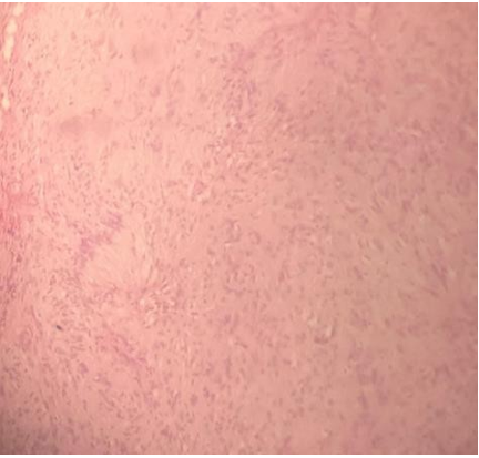
Figure 6: Photomicrographs of the Antoni B pattern.
The patient has not shown any recurrence in follow-up period of 1 year.
Schwannoma was first described by Verocay in 1910. Schwannomas are benign encapsulated nerve sheath neoplasm composed of Schwann cells [5]. Wright and Jackson reported 146 cases of schwannoma of the oral cavity soft tissue. Of those, 52% occurred in the tongue, 19.86% in the buccal or vestibular mucosa, 8.9% in the soft palate, and the remainder 19.24% in the gingivae and lip [6]. It is relatively an uncommon neural neoplasm with unknown etiology [7]. Malignant transformation is rare and incidence of malignant schwannomas ranges from 8% to 13.9% [8].
In embryological development, Schwann cells originate around the fourth week from a specific group of ectomesenchymal cells derived from the neural crest. These cells act as a protective layer around each peripheral nerve fiber of both motor and sensory nerves. Additionally, they encase larger nerve fibers with a myelin sheath, which enhances the speed of nerve signal transmission [9].
Neurilemmomas occurring inside the mouth are rare, accounting for only 1% of all oral cavity tumors. Their uncommon occurrence can result in initial misdiagnosis during oral lesion assessments. This underscores the importance of conducting a comprehensive and meticulous evaluation when diagnosing and treating oral conditions. Rare conditions like neurilemmomas present distinct challenges and often require specialized management approaches [10].
Diagnostic investigations typically encompass ultrasound scanning, computed tomography (CT), magnetic resonance imaging (MRI), and fine-needle aspiration cytology. Standard histopathological examination provides a definitive diagnosis of schwannoma. The tumor tissue exhibits distinct Antoni A and B regions: Antoni A areas are characterized by densely packed spindle cells arranged in a palisading pattern around acellular eosinophilic areas known as Verocay bodies, formed by thin cytoplasmic fibers and duplicated basement membranes. In contrast, Antoni B areas tend to have a more myxoid consistency. Additionally, the tumor tissue may show features such as hemorrhage from adjacent tissues, necrosis, hyalinization, and cystic degeneration.
The primary clinical differential diagnoses for other benign neoplasms at this location include neurofibroma, traumatic neuroma, nonossifying fibroma, lipoma, and leiomyoma [11].
The histopathological variants of schwannoma are ancient schwannoma, pseudoglandular schwannoma, plexiform schwannoma, cellular schwannoma, epithelioid schwannoma, and melanotic schwannoma. Woodruff et al in 1981 reported a series of 14 cases of cellular schwannomma that were histologically characterized by increased cellularity, nuclear pleomorphism, hyperchromatism, lack of verocay bodies, and frequently higher mitotic activity [12].
Treatment for schwannomas typically involves surgical excision, which is the primary therapeutic approach. The goal of surgery is complete removal of the tumor while preserving nerve function as much as possible. This is especially crucial in cases where the tumor is located near important nerves or structures.
Here are some key points about treatment
Overall, the prognosis for schwannomas is generally good, especially with complete surgical removal. Recurrence rates are low after successful surgery, and most patients experience relief from symptoms associated with the tumour.
Till date, very few cases have been reported in the oral cavity. Its incidence is about 1% in the oral cavity, making it even more unusual in the alveolar mucosa in association with the mental nerve.
Department of Oral Pathology, HIDS, Paonta Sahib.
None.
Copyright: © 2024 Vanshikha Sharma., et al. This is an open-access article distributed under the terms of the Creative Commons Attribution License, which permits unrestricted use, distribution, and reproduction in any medium, provided the original author and source are credited.
ff
© 2024 Acta Scientific, All rights reserved.