Mithun J Shah1*, Kenneth FH Tan2, K Sridharan3, Naveen Kumar Krishna4 and Nagarathna KN5
1Orthodontic and Dentofacial Orthopeadic, Reader, Department of Orthodontics and
Dentofacial Orthopeadics, KGF College of Dental Sciences and Hospital, Karnataka, India
2Orthodontics and Dentofacial Orthopaedics, Formerly Professor and HOD, Department of Orthodontics and Dentofacial Orthopaedics, The Oxford Dental College and Hospital,
Karnataka, India
3Orthodontic and Dentofacial Orthopeadics, Professor and HOD, Department of Orthodontics
and Dentofacial Orthopeadics, Sri Siddhartha Dental College and Hospital, Karnataka, India
4Orthodontic and Dentofacial Orthopeadic, Professor and HOD, Department of Orthodontics
and Dentofacial Orthopedics, KGF College of Dental Sciences, Karnataka, India
5Orthodontic and Dentofacial Orthopeadic, Department of Orthodontics and dentofacial Orthopaedics, KGF College of Dental Sciences, Karnataka, India
*Corresponding Author: Mithun J Shah, Orthodontic and Dentofacial Orthopeadic, Reader, Department of Orthodontics and Dentofacial Orthopeadics, KGF College of Dental Sciences and Hospital, Karnataka, India.
Received: August 28, 2024; Published: September 26, 2024
Citation: Mithun J Shah., et al. “Load Deflection Characteristics of Different NITI Archwires with Modified Bending Test Simulating Clinical Situation-An In vitro Study". Acta Scientific Dental Sciences 8.10 (2024):52-62.
Background: Orthodontic archwire is critical in orthodonic treatment as superelasticity, shape memory, and the capacity to provide continuous force across a wide range of action, optimal archwire, superelastic NiTi, and Heat Activated NiTi (HANT) archwires have been gaining popularity as initial alignment archwires.
Aims and Objectives: To investigate the load deflection characteristics of HANT and superelastic NiTi wires in the latest commonly used MBT prescription in both ligating and self-ligating brackets with modified bending test simulating a standardized arch form with the aim of selecting the most appropriate aligning wires.
Materials and Methods: Acrylic full arch form models were fabricated using 19 x 25 upper stainless steel archwire with portion holding the maxillary left lateral incisor bracket was free to move in vertical and horizontal application of forces. Each arch wire specimen was inserted into acrylic model with the lateral incisor region of the archwire engaged and activated to the designated distance of (5, 4, 3, 2 and 1 mm). Each bending test was carried out 3 times with a new piece of archwire for each repetition.
Results: In occluso-gingival direction, VSB showed binding effect above 3mm deflection on HANT wire and 4mm deflection on Superelastic wire. In labio-palatal direction, VSB showed binding effect above 4mm deflection on HANT wire and no binding was seen on SE wire. Further, no binding effect was seen on SLB at any point of deflection on HANT and SE wires. So HANT and SE wire can be used on SLB in both occluso-gingival and labio-palatal direction up to 5mm deflection.
Conclusion: We conclude that SE and HANT wire on SLB showed better performance when compared to VSB in both occluso-gingival and labio-palatal direction in initial aligning stage.
Keywords: Superelastic; Load Deflection Point; Nickel Titanium; Heat Activated Nickel Titanium
The decision regarding the use of an orthodontic archwire is critical in orthodonic treatment. For every single person, this decision ought to be determined by estimated forces, which are the best force for the highest effective tooth displacement. Because of its unique qualities of superelasticity, shape memory, and the capacity to provide continuous force across a wide range of action, optimal archwire, superelastic NiTi, and Heat Activated NiTi (HANT) archwires have been gaining popularity as initial alignment archwires [1-3].
However, knowledge about the actions of these wires has its foundation on typical three-point bending tests, [4] which do not simulate the variability seen in clinical situations, despite their reliability [5].
The researchers employed straight-length wires without taking archform into account or mimicking the clinical situation of loading in the occluso-gingival and labio-palatal directions, which are frequent during alignment.
As a result, a suitable three-point bending test must recreate clinical conditions, with wires confined to the vicinity of fixed appliances [6]. Studies have also found that variations in the model architecture employed for such tests appear to alter the loading-unloading graph as well as should be taken into account when assessing data [7].
Therefore, the purpose of this study is to investigate the load deflection characteristics of HANT and superelastic NiTi wires in the latest commonly used MBT prescription in both ligating and self-ligating brackets with modified bending test simulating a standardized arch form with the aim of selecting the most appropriate aligning wires.
Two 0.016-inch round superelastic and heat activated NiTi archwires on two different 0.022 x 0.028-inch MBT prescription bracket system was tested in-vitro to compare their mechanical properties.
2 acrylic full arch form models were fabricated using 19 x 25 upper stainless steel archwire, one for the conventional victory series brackets and the other for the self-ligating smart clip brackets. The portion holding the maxillary left lateral incisor bracket was free to move in vertical and horizontal application of forces, in the model.
To simulate intraoral clinical situation, maxillary 0.022 x 0.028-inch stainless steel brackets and first molar tubes were bonded to the lateral surface of acrylic plate using self-cure acrylic resin and adhesive with no angulations/torque in the system.
The inter-bracket distance was derived from typical tooth dimensions (accurate to within 0.5mm) for a maxillary permanent dentition. The brackets were positioned in accordance with the average tooth width to simulate full arch form (with their long axis parallel to the acrylic arch form model).
In model 1 with conventional victory series brackets were bonded with 3M elastomeric power modules ligature was used to engage arch wires which were placed with a straight shooter ligature gun to minimize the variation in the degree of stretching during application and in model’ 2’ with self-ligating smart clip brackets, arch wires were engaged using the smart clip inserting tool
The acrylic jig with the wire specimens was suspended in an artificial saliva bath (37 ±1° C) in polymethyl methacrylate box measuring (4”x 4” x 4”). Each arch wire specimen was inserted into acrylic model with the lateral incisor region of the archwire engaged and activated to the designated distance of (5, 4, 3, 2 and 1 mm).
Deactivation force was measured using a instron universal testing machine (Model 5563) in with a 100 kg compression load cell set on a range of 2 kg operating at a crosshead speed of 1.0mm/minute and deflection rod tip formed from 6 mm diameter chisel attached to upper chamber of instron machine having load cell.
Each bending test was carried out 3 times with a new piece of archwire for each repetition.
The wires were loaded in a labio-palatal direction and occluso-gingivally in the arch form model to allow greater stability and positional accuracy. Wires were deflected to 1, 2, 3, 4, and 5 mm in separate deflection test.
Load value measurements from the unloading phase were derived digitally by selecting 2 deflection points on an x - y plot on the computer screen (x axis - compression extension and y axis as compression load) which was auto calibrated. Few points were chosen to represent a standardized portion of the unloading phase plateau in the load-deflection curve.
For test deflection at 1mm, the load values were measured at 0.5 mm, for remaining, the deflection point chosen was 0.5mm less than the maximum deflection of each bend and 1.0mm.
Mean and standard deviation of the forces generated at activation and deactivation for a deflection of 1,2,3,4 and 5 mm were calculated with data for the deactivation force analyzed by student “t” test
Most but not all, load deflection graphs of the Super elastic NiTi and HANT wires confirmed features of super elasticity, with plateau regions varying in extent, gradient, and load value depending on wire, model, temperature and method of attachment.
A mean load value is given for each of the 2 points on the curve, and the plateau gap is the average load difference between initial and final unloading deflection points. It therefore represents a measure of gradient of unloading plateau over a standardized deflection distance and small proportional change in force level.
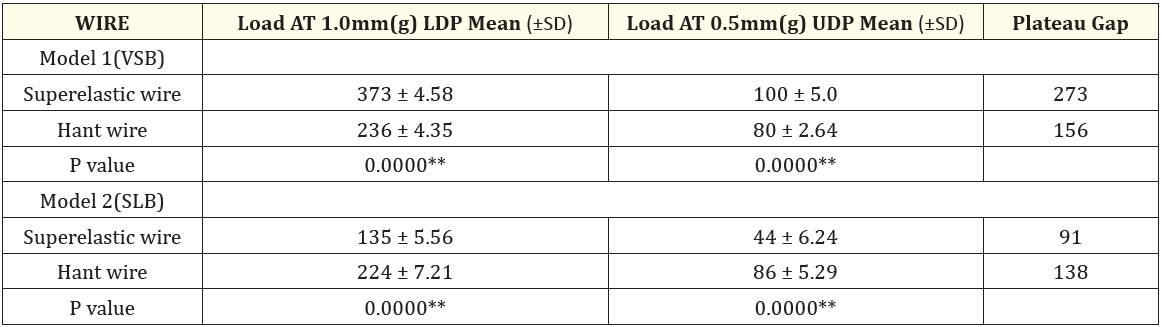
Table 1: Comparision of the wires on model 1 and model 2 in occluso-gingival direction at 1mm deflection.
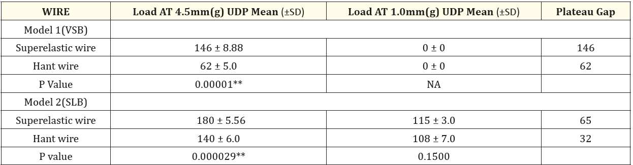
Table 2: Comparision of the wires on model 1 and model 2 in occluso-gingival direction at 5 mm.
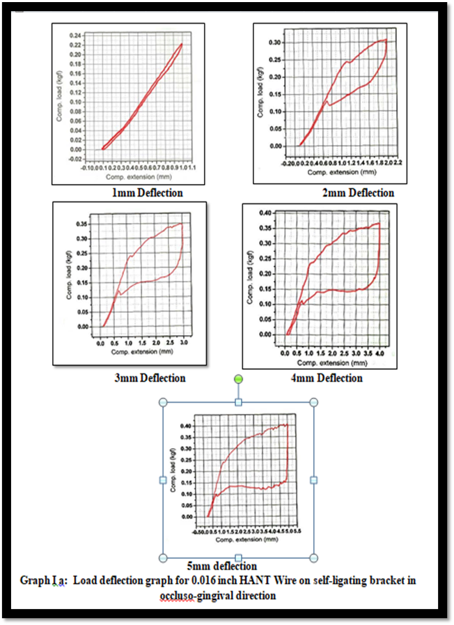
Figure 1
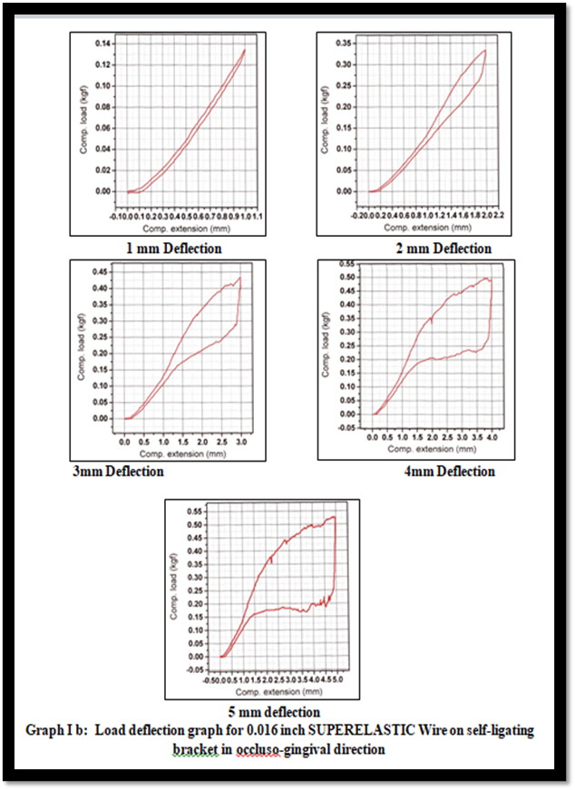
Figure 2
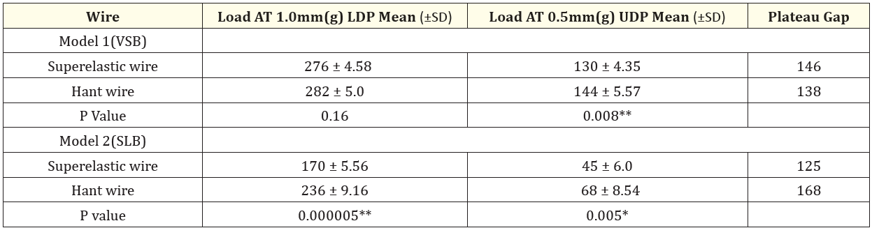
Table 3: Comparision of the wires on model 1 and model 2 in labio-palatal direction at 1 mm.
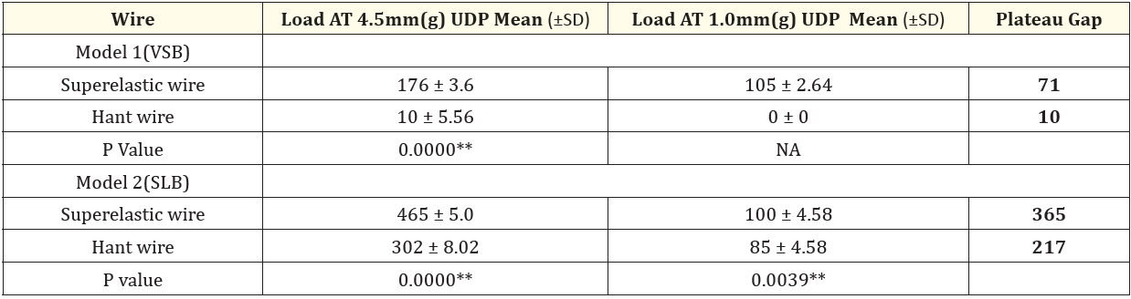
Table 4: Comparision of the wires on model 1 and model 2 in labio-palatal direction at 5 mm.
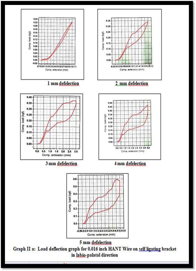
Figure 3
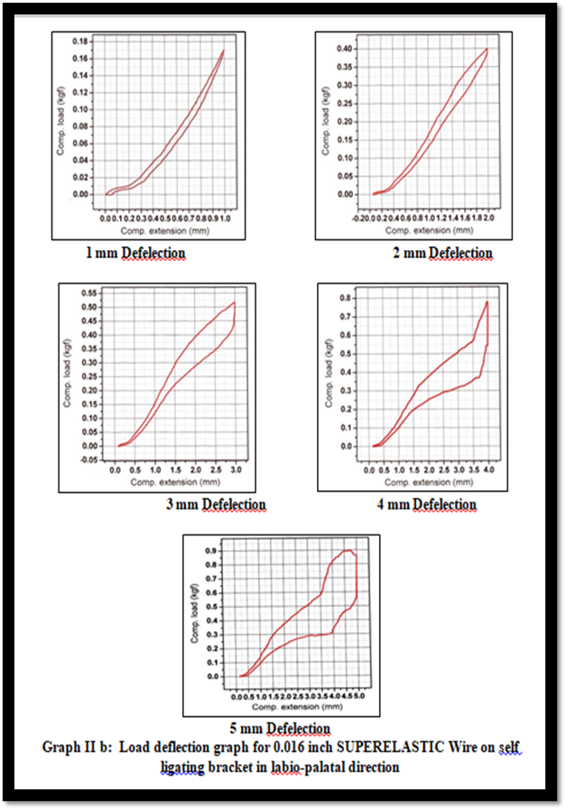
Figure 4
The subject of optimal orthodontic forces has been continually discussed since the early 20th century. Schwarz defined ideal force as that which causes a pressure shift similar to capillary pressure, hence preventing vascular occlusion in the compressed area of the periodontal ligament. This hypothesis has been restated by authors including Reitan, Righ., et al, and Proffit, but no research has yet been conducted to measure this optimum force when applied continuously [8].
Nowadays, ''light continuous forces'' are regarded to be physiologically acceptable and efficacious, and producing such a force system is the major problem in creating and operating an orthodontic device [8].
Superelasticity is a unique feature that results from stress-induced martensitic transformation. When an external force is applied to the Ni-Ti wire, it deforms by martensitic transformation rather than lattice sliding, as is prevalent in most metals. During this phase, the metal becomes increasingly soft and ductile. When stress is reduced, the Ni-Ti alloy reverts to the harder austenitic phase.
Clinically, the superelasticity enables the archwires to produce a consistent force across a wide range of activities. It does not behave linearly elastically, but rather generates continuous force over lengthy periods of time.
The comparison of the mechanical properties of the NiTi superelastic wires and wires of other alloys presents outstanding differences; the structural properties (form and dimension) seem to have a similar outcome in the movement of teeth with regard to deflection.
According to Burstone, Koenig, Waters and Hemingway et al many complex factors influence the performance of an orthodontic archwire in the mouth. These factors are as follows
The explanation for this could be frictional resistance throughout the wire and bracket slot, or force fluctuation amongst the unrelaxed elastomerics utilized, and the results were comparable to Wilkinson PD [1,4].
When compared to VSB, the plateau gap reveals a consistent delivery of force on SLB on SE and HANT wire, implying that it can be employed in mild to moderate crowding situations. As the degree of three-point deflection grows to 2mm, the slope of the unloading curve becomes significantly reliant on both the initial deflection and the extent of recovery.
The VSB and SLB demonstrated statistically significant results for unloading SE and HANT wire at 1.5mm, with a linear graphic depiction in the occluso-gingival and labio-palatal directions. VSB gave the shortest load values on discharging at 1.5mm deflection, demonstrating that more frictional resistance between these brackets and the wire decreased the load values when compared to SLB, which caused substantially less friction, and the wire were therefore able to exert higher loads [1,9].
According to Bartzela TN, Senn C, and Wichelhaus, a narrower plateau gap during the unloading phase indicates a lower and more steady force in clinical applications. Derotational operations require a wire with a longer clinical plateau and larger application force [10].
In our study, the HANT wire had the smallest plateau gap on SLB in the occluso-gingival direction and the smallest plateau gap on VSB in the labio-palatal direction, indicating a low and consistent force in those directions. When compared to SE wire, these data indicate that HANT wire on SLB can be utilized when teeth are strongly positioned, while HANT wire on VSB can be used when teeth are mildly to moderately crowded.
The 3mm three-point deflection revealed statistically significant results for all wires and brackets in both directions. A linear graph for HANT wire on SLB and VSB in the occluso-gingival direction, HANT and SE wire on SLB in the labio-palatal direction, and the rest curve displayed "Steps and Spikes" graphically. This was due to slippage of the wire inwards over the supports as the deflection rose, and the spike in the graph was caused by a sudden rise in force, which could indicate that the wire was completing its reverse transformation back to the austenitic metallurgic phase [9].
On unloading to 2.5mm, SLB provided higher load than VSB on both SE and HANT wire in both directions, and the cause was because of less frictional in the wire.
The plateau gap revealed an unusual behavior on SE and HANT wires on VSB in the labio-palatal direction, with negative values.3 According to Mallory DC, this was owing to a lack of sufficient heat to return the wire to its totally austenitic phase. And HANT wire on SLB created the smallest plateau gap of any wire, showing the least and most consistent force, implying that these wires can be employed in mild to moderate crowding in both directions.
At 4mm three-point deflection, there was a statistically significant difference in 3.5mm unloading in the occluso-gingival and labio-palatal directions compared to 1mm unloading. The entire graph began to display "Steps and Spikes" in graphic depiction, with the exception of the HANT wire on SLB in occluso-gingival and labio-palatal directions and the SE wire on SLB in labio-palatal direction.
When unloaded to 3.5mm deflection, VSB showed less load than SLB, however when unloaded to 1mm deflection, the wire did not restore to its previous form due to the binding effect seen. This demonstrates that the SE or HANT wire performed poorly when used on VSB with deflections greater than 3mm due to the binding effect. The plateau gap was negative on HANT wire in VSB in the labio-palatal direction, and the smallest plateau gap was detected on HANT wire on SLB in the occluso-gingival direction, indicating that it can be employed in strongly placed teeth [3].
At 5 mm three-point deflection, there was a statistically significant effect in 4.5mm unloading in the occluso-gingival and labio-palatal directions, but not at 1mm unload. Except for the SE and HANT wire on SLB in the labiopalatal direction, all graphs displayed "Steps and Spikes" graphically. When unloaded to 4.5mm deflection, VSB showed less load than SLB, however when unloaded to 1mm deflection, the wire did not restore to its previous form due to the binding effect seen. This demonstrates that the SE or HANT did not function well on VSB with deflections greater than 3mm due to the binding effect. The least plateau gap was observed on SE and HANT wire on SLB in the occluso-gingival direction, indicating that it can be employed in highly placed teeth.
Superelastic wire responds mechanically very differently from HANT wire on different brackets system. After the wire is deflected from 1-5mm and deactivation occurs, wire-slot friction at the support reduces the net unloading force at the lateral incisor bracket as the deflection increases. These changes in frictional and spring back forces occurred for two reasons.
First, frictional force creates wire and bracket-slot contacts at the central incisor along with canine brackets. As the deflection amplitude grows, so does the wire curvature between the lateral incisor and the supporting bracket slots, which in turn curvature leads in higher normal and frictional forces at the supportive bracket slots. Second, increased deflection-amplitude with SE and HANT wire resulted in lower unloading stiffness and spring back forces. A deflection condition may occur when the wire's spring back potential is insufficient to counteract frictional forces, causing the wire to bind in those slots.
In the occluso-gingival direction, VSB demonstrated binding above 3mm deflection on HANT wire as well as 4mm deflection on SE wire. In the labio-palatal direction, VSB demonstrated binding over 4mm deflection on HANT wire, and no binding was seen on SE wires.
Binding was stronger in the occluso-gingival direction than in the labio-palatal direction because SE wires had a higher loading force than HANT wires, and vice versa.
When viewed clinically, a bound wire may "free-up" and unload as a result of the coupling exerted, which may twist the central incisor and canine towards the malpositioned lateral incisor, resulting in either no tooth movement or undesirable tooth movement. It should be observed that SE and HANT wires bind in both the occluso-gingival and labio-palatal directions.
Some studies suggest that self-ligating brackets create a "virtually friction-free" appliance in which binding is impossible [11,12].
The unloading curves in this study fell within limited boundaries, despite varying degrees of superelastic activity. The unloading plateau occurs at a clinically meaningful displacement of 1 to 2.5 mm. This unloading plateau region is the force value that is most likely to be used in a clinical setting once teeth have moved inside the periodontal ligament.
We discovered that the load deflection performance of 0.016-in alignment wires varied based on the test model's design, bracket type, and degree of deflection. In addition, HANT wire exhibited the lowest and constant deactivation force, while SE wire had the largest deactivation force on unloading in both VSB and SLB in the occluso-gingival and labio-palatal directions.
Therefore, we advocate a multi-centric, large sample, randomized-controlled, standardized study to eliminate all the shortcomings encountered in our study.
We found that SE and HANT wire on SLB showed better performance when compared to VSB in both occluso-gingival and labio-palatal direction in initial aligning stage. We recommend further investigation required to address other aspects like biological resistance and bracket angulations, so this test may have a limited application to clinical conditions.
Copyright: © 2024 Mithun J Shah.,et al. This is an open-access article distributed under the terms of the Creative Commons Attribution License, which permits unrestricted use, distribution, and reproduction in any medium, provided the original author and source are credited.
 Open Access by
Acta Scientific is licensed under a Creative Commons Attribution 4.0 International License
Open Access by
Acta Scientific is licensed under a Creative Commons Attribution 4.0 International License
Based on a work at https://actascientific.com
ff


ff
© 2024 Acta Scientific, All rights reserved.