Hemalatha DM1*, Subair K2, Vishnu Sripriya1, Liyana Hassan3 and Megha RP3
1Assistant Professor, Department of Periodontics, Mahe Institute of Dental Sciences, Chalakkara, India
2Professor, Department of Periodontics, Mahe Institute of Dental Sciences, Chalakkara, India
3Third Year BDS Student, Mahe Institute of Dental Sciences, Chalakkara, India
*Corresponding Author: Hemalatha DM, Assistant Professor, Department of Periodontics, Mahe institute of Dental Sciences, Chalakkara, India.
Received: August 09, 2024; Published: August 28, 2024
Citation: Hemalatha DM., et al. “Artificial Intelligence in Dentistry- A Review". Acta Scientific Dental Sciences 8.9 (2024):87-91.
Artificial intelligence has become ubiquitous in day to day activities. Its use is inevitable and the medical profession has started to use the mimicking use of artificial intelligence in complex proposed predictions and decision making. AI have shown numerous applications in dentistry predicting the wide variant anatomy of the jaws. hese days, a computer can identify or predict a result from a large database thanks to machine learning. The AI based comprehensive care system is anticipated to establish superior patient care as well as creative research and development in the future, enabling cutting edge decision support tools.
Keywords: Neural Network; ANN; CNN; Artificial Intelligence
John McCarthy created the term artificial intelligence (AI), which describes machines that can mimic human knowledge and behavior. The term artificial intelligence (AI) has gained widespread usage due to its excessively broad definition. One area of AI that academics and industry professionals have extensively used for data analysis is machine learning. Arthur Samuel originally discussed machine learning in 1959, defining it as a method that allows computers to learn without explicit programming. T AI will continue to have a significant impact on dentistry from a broad standpoint because of the necessity for accurate treatment methods and immediate information interchange, even in the face of potential misunderstandings and patient privacy concerns [1].
AI technologies have also been widely adopted in the medical field, primarily in the area of computer thus far. Diagnostic imaging is essential and inevitable in healthcare. Artificial intelligence (AI) is particularly well-suited to address the variability in subjective individual examinations and to improve care effectiveness.
AI makes the best use of multilevel data and enables an understanding of interactions by integrating heterogenous data domains, such as medical/dental history, socio-demographic and clinical data, imaging data, biomolecular data, socio-network data, etc. AI has the potential to improve mundane tasks and boost in-person interaction between medical professionals and their patients (also known as “Humanizing Care”).
AI also promises to increase patient participation in healthcare, particularly if individuals actively contribute their data through other means. Self- management and self-monitoring will empower. In order to support the world health organization’s (WHO) sustainable development goals, AI may also help alleviate human resource or manpower shortages, which have been noted and are predicted to persist in many regions of the world [2].
The last 20 years have seen a significant advancement in AI research, and it is hard to find a setting where AI is not used in some way in daily or work-related task requiring digital or mobile devices. It is imperative that the machine run in order for this to happen. Understanding how AI is used in daily life would be helpful given the general trend of AI [3].
ANN is a simple deep learning model that consists of three layers . It is made up of a bunch of neurons and layers. Only forward processing of the input is done. From the input layer, input neurons extract features from the data and send it to hidden layers, where it passes through each hidden layer in turn. The output layer displays a summary of the final results. Before forwarding the data to the following layer, each hidden layer in an ANN has the ability to balance the data received from earlier layers and make adjustments. The ANN can comprehend more complicated features since each hidden layer serves as an input and output layer [4].
The primary applications of CNN, a kind of deep learning model, are in picture production and recognition. The pooling layer, fully connected layer, and convolution layers are additional components of CNN's hidden layers, which is the main distinction between it and ANN. Using convolution kernels, convolution layers are used to create feature maps from input data.
Convolutional weight sharing results in a reduction of visual complexity. In order to lower the dimension of feature maps for additional feature extraction, each group of convolution layers typically comes after the pooling layer. The pooling and convolution layers are Convolutional employed before the fully linked layer. The completely connected layer, as its name suggests, converts 2D feature maps into 1D by connecting to every neuron that was active in the preceding layer. After that, nodes of categories are linked to maps in order to classify them. When compared to ANN, CNN demonstrated greater efficiency and accuracy in image identification [4].
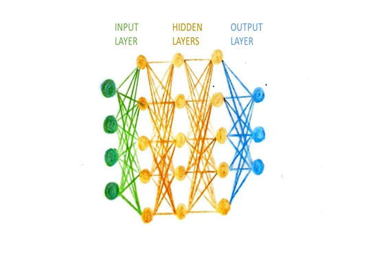
Figure 1: Growth Parameters of Chilli under foliar application of treatments
Periodontitis is one of the most common disease. It affects billions of people and, if left untreated, it can result in tooth loss and/or mobility. Early detection and treatment of periodontitis are necessary to prevent severe cases. In clinical practice, gingival recession and pocket probing depths are used to diagnose periodontal disease. Clinical attachment loss is commonly quantified using the Periodontal Screening Index (PSI). This clinical evaluation, however, is not very reliable because dentists' experience still plays a major role in the screening process for periodontal disease, which may cause them to overlook localized periodontal tissue loss. Artificial intelligence has been applied to periodontics to diagnose periodontitis and categorize potential periodontal disease types. Krois., et al. also used CNN to identify periodontal bone loss on panoramic radiographs. The accuracy and potential utility of a suggested CNN algorithm to automatically identify teeth with periodontal deterioration were assessed by Lee., et al. Yauney., et al. asserted that a CNN algorithm created by their study team using information about systemic health could assess periodontal diseases [4].
In the context of machine learning, a sufficient amount of data is needed to train and improve an AI tool. It is acknowledged that supervised training is essential to raising a CNN's effectiveness. In order to establish structures for the CNN to segment, this method necessitates that tagged images be displayed to the CNN. Nonetheless, overtraining CNNs can lead to over recognition and mistakes [8].
Luciano., et al. developed Haptics-based virtual reality periodontal training simulator. Students can learn the skills needed to diagnose and treat periodontal disorders with the aid of this simulator. In 1998, Companion., et al. first published the results of an ultrasonographic periodontal probe at NASA Langley. The goal of this probe was to lessen the discomfort and error that come with manual probing. In 2017, Feres., et al. tested a hypothesis by utilizing a linear Support Vector Machine (SVM)-based classifier and 40 bacterial species of the subgingival microbial complexes to distinguish between Generalized Aggressive periodontitis in younger adults and Generalized Chronic periodontits.
A machine learning classifier that could differentiate between inflamed and healthy gums was published by Rana, Yauney., et al. in 2017. An oral imaging device was used to record the biomarker porphyrin's associated fluorescence upon exposure to light with a wavelength of 405-450 nm. Plaque was shown in yellow and orange tones, whereas red and magenta tones indicated inflamed gums.
A methodology for employing AI to fabricate implant-supported monolithic zirconia crowns (MZCs) cemented to separate hybrid abutments was presented by Lerner, Mouhyi., et al. in 2020. Here, the crown was created using CAD software and AI. The benefits included saving time, lowering the risk of mistakes, and lowering the expense of prosthetic rehabilitation [7].
Artificial Intelligence is still relatively new to the field of periodontology and implantology, and its full potential has not yet been realized. Applying this tool seems to have a lot of potential benefits because of its advantages in data analysis, detailed regression, and diagnostic aid. Compiling databases is a problem in any data research. This presents a serious challenge for gathering information from medical records.
Reliable predictions have been produced by developing AI algorithms for Periodontal risk assessment and comparing them with earlier models. Since these AI algorithms were easy to understand and considered a range of periodontal disease-related characteristics, it has been discovered that they are straightforward yet useful tools for making decisions when predicting periodontal disease.
Radiographic bone loss (RBL) calculations can be difficult, time-consuming, and examiner-specific. Thus, artificial intelligence (AI) algorithms have been developed to automatically detect RBL as well as the risk of periodontal disease and tooth loss. The ability of AI models to identify RBL for the diagnosis of periodontal disease was examined by Miller., et al. [2023]. They found that periapical radiographs demonstrated a precision of 25% for detecting moderate disease and a high accuracy of 99% for staging RBL, whereas the mean accuracy for panoramic radiographs ranged between 63% and 94%. On cone-beam computed tomography (CBCT), the specificity for periodontal bone loss varied from 81% to 83%, whereas the sensitivity was 45-72%.
For artificial olfaction, the entire spectrum of inhaled volatile chemicals is assessed using a variety of non-selective sensors. Because it combines artificial intelligence (AI) with mammalian olfaction to recognize particular scent patterns and serve as a reference for subsequent identification, it is also known as an electronic nose. The programmed sensor array is divided into two subsets: the top panel is more suited for non-sulfuric volatile organic molecules, while the bottom panel is more suited for volatile sulfur compounds (VSCs). The sensors respond in unison when exposed to the sample, and their responses are analyzed to look for patterns. The software makes a comparison between the patterns gathered from various sensors and the database of patterns acquired earlier in the preclinical training stages. If a subject has extraoral or oral halitosis, it is determined by a decision tree-based classifier. Additionally, it will categorize the volatolomic pattern in the event of extraoral halitosis according to several systemic disorders [9].
According to a certain set of criteria, dentists have historically diagnosed caries by visual and tactile examinations or radiographic examinations. When deep fissures, tight interproximal contacts and secondary lesions are present, it can be difficult to detect lesions in the early stages. A Research has been done in the field of operative dentistry on the identification of dental caries, apical lesions, vertical root fractures, pulp space volumetric assessment, and tooth wear evaluation. In a two-dimensional (2D) radiograph, the density of the item is represented by the brightness of each grayscale picture pixel. An AI program may identify patterns and make predictions to segment teeth, identify cavities, and other tasks by analyzing the above mentioned features. A CNN algorithm was created by Lee., et al. to identify dental caries on periapical radiographs. A CNN method was presented by Kühnisch., et al. to identify dental caries on intraoral pictures. When Schwendicke., et al. compared the cost-effectiveness of artificial intelligence (AI) with dentist’s diagnosis for proximal caries detection, the findings indicated that AI was more efficient and less expensive. According to a number of the previously mentioned research, artificial intelligence (AI) exhibits the same or even greater accuracy in early lesion diagnosis than dentists [4].
The experience and preferences of the orthodontists are typically taken into consideration while planning orthodontic treatment. It has historically taken a lot of work for orthodontists to identify malocclusion since a cephalometric analysis must take into account a number of variables, making it challenging to decide on a course of therapy and forecast its success. AI is used in treatment planning and outcome prediction. For example, it can simulate how pre- and post-treatment facial photos will seem different. With the use of AI algorithms, patients and dentists may communicate more effectively by seeing the effects of orthodontic therapy, skeletal patterns, and anatomic landmarks in lateral cephalograms.
To assess if extractions from lateral cephalometric radiographs are necessary, Xie., et al. suggested an artificial neural network (ANN) model. A high-accuracy DL method for automatically recognizing cephalometric landmarks on radiographs was presented by Park., et al. Segmenting and categorizing the teeth is a fundamental process for orthodontic treatment planning. These objectives have also been met by AI on a variety of sources, including radiography and full-arch 3D digital optical scans. A number of AI methods were proposed by Cui., et al. to automatically segment teeth on a digital dental model that was scanned using CBCT images and a 3D intraoral scanner [4].
Oral cancer is the most serious form of OMFP. The main application of AI research has been the detection of tumors and cancer using radiography, microscopy, and ultrasonography images. AI is also capable of identifying anomalous areas from radiographs, including the salivary and parotid glands, interdigitated tongue muscles, and oral cavity nerves. AI is used in pre-surgical orthopaedics, speech evaluation, surgery, risk assessment, and diagnosis while treating cleft lip and palate.
In order to identify oral squamous cell carcinoma (OSCC) and oral potentially malignant disorders (OPMDs) in intraoral optical images, Warin., et al. employed a CNN technique. James., et al. distinguished between malignant and dysplastic oral lesions using ANN and SVM models. Abureville., et al. automatically diagnosed oral squamous cell carcinoma (SCC) from confocal laser endomicroscopy pictures using a CNN algorithm. A CNN algorithm was utilized by Poedjiastoeti., et al. to detect and differentiate between ameloblastoma and keratocystic odontogenic tumor [4].
In prosthodontics, preparing a tooth, taking an impression, trimming the cast, designing, fabricating, trying in, and cementing the restoration are all common steps in the construction of a dental crown. Digital design work has been made possible by CAD/CAM in commercially available solutions like 3Shape, Sirona, and CEREC. By using a tooth library for crown design, this has significantly improved the design process' efficiency, but it still falls short of producing a unique design for each patient. Hwang., et al. and Tian., et al. suggested cutting-edge methods based on 2D-GAN models to create a crown by learning from technicians' ideas as AI developed. 2D depth maps created from 3D tooth models served as the training set.
AI has also been applied to the prediction of debonding and shade matching in CAD/CAM restorations. Ding reported a 3DDCGAN network in the crown generation, which utilised 3D data directly in the crown generation process, the morphology of generated crowns was similar compared with natural teeth. In contrast to fixed prosthodontics, removable prosthodontics requires more consideration of variables, making design more difficult. There isn't a machine learning method available for creating detachable dentures, but there are a number of expert systems that rely on knowledge. Present-day machine learning algorithms are primarily aimed at supporting the design of removable dentures. For example, they classify denture arches and predict edentulous patients based on their facial features [4].
Artificial intelligence (AI) has become a potent instrument in the field of smile design, capable of improving treatment planning's precision and effectiveness and, eventually, improving patient results. By recommending the best course of action based on accepted dentistry principles and rules, AI can help automate the smile design process. This lowers the possibility of human error while also saving dentists time. The technically challenging treatment process of Digital Smile Design can be completed more easily with the help of software that has been upgraded with artificial intelligence. In several areas of dentistry, the usage of Digital Smile Design (DSD, smilecloud 2.0, Rubicon (2020)) approaches has increased. A computer or laptop, intra- and extra-oral pictures of the patient, and a general understanding of dental aesthetics are needed to create a smile design. Since the protocol uses an intra-oral scanner rather than a traditional impression, the patient may perceive the procedure as being easier, more convenient, and less painful. A cloud-based platform called SmileCloud (Version 2.0, Rubicon) enables the online storage of all patient data, including pictures, intra-oral scanning, cone beam computed tomography (CBCT), and functional mandibular movement, without any space restrictions or data loss potential [5].
The increasing prevalence of AI-enabled work applications requires the attention of occupational safety and health practitioners, researchers, employers, and employees. Occupational safety and health professionals must have strategic foresight to predict and plan for the opportunities and difficulties of AI-enabled technologies on worker safety, health, and well-being in order to take a proactive approach to AI and its implications for the future of work [6].
Artificial Intelligence (AI) has brought about changes in the healthcare sector, attracting investment from both corporations and researchers. Dentistry, and dental research in particular, has a responsibility to guarantee that AI will improve dental treatment at a reduced cost for the benefit of patients, clinicians, and society at large.
Copyright: © 2024 Hemalatha DM., et al. This is an open-access article distributed under the terms of the Creative Commons Attribution License, which permits unrestricted use, distribution, and reproduction in any medium, provided the original author and source are credited.
 Open Access by
Acta Scientific is licensed under a Creative Commons Attribution 4.0 International License
Open Access by
Acta Scientific is licensed under a Creative Commons Attribution 4.0 International License
Based on a work at https://actascientific.com
ff


ff
© 2024 Acta Scientific, All rights reserved.