Nandlal B1, Karthikeya Patil2 and Ravi S3*
1Dean Faculty of Dentistry, JSS University & Professor in Pedodontics and Preventive Dentistry, JSS Dental College and Hospital, Karnataka, India
2Professor & Head Department of Oral Medicine & Radiology, JSS Dental College and Hospital, Karnataka, India
3Associate Professor, Department of Orthodontics, JSS Dental College and Hospital, Karnataka, India
*Corresponding Author: Dr. Ravi S, Associate Professor, Department of Orthodontics, JSS Dental College and Hospital, Mysore, Karnataka, India.
Received: May 22, 2017; Published: June 05, 2017
Citation: Ravi S., et al. “Craniofacial Changes in Rural Residential School Children of Suttur of 7 Years of Age”. Acta Scientific Dental Sciences 1.1 (2017): 09-16.
Introduction: Craniofacial growth is a complex and beautiful phenomenon. Growth has been described in so many terms. Todd defines growth as “increase in size”. Meredith defines growth as the entire series of anatomic and physiologic changes taking place between the beginning of prenatal life and the close of senility. Growth is quantitative, i.e., it is measureable aspect of biologic life.
Objective: The present study was designed to describe the craniofacial dimensions of rural residential free school children of Suttur at 7 years.
Material and Methods: In order to study the changes in Cepholometric measurements 10 Males & 10 Females of 7 years old children were selected and Lateral Cephalograms were taken. On each Lateral Cephalogram, 21 skeletal cephalometric landmarks were identified. From the cephalometric landmarks and reference lines, 31 angular and linear measurements were analyzed, which were grouped as JSS Craniofacial analysis.
Results & Discussion: Mean Sagittal Measurements of 7 year old, when compared between males and females, the females showed a higher values than males for the variables SNA, SNB, ANB, A-Npog, A:ss-n-pg. Mean Basal Vertical measurements of 7 year old when compared between Males and females for values of PFH, PFH/AFH, Jaw relation, Maxillary Inclination, Jaw angle, Mandibular Inclination, SN-PoOr & ANS: Gn there was no statistically significant difference between Males and Females. Mean Mandibular length measurements Go-Co and Co-Gn of 7 year old when compared between males and females, the males had higher Mean values than the females for both the parameters. The Mean cranial base measurements of 7 year old when compared between Males and females, the males had higher Mean values than the females for S:Ba, PCB & ACB, but females had higher mean values than males for n-s-ar & n-s-ba.
Conclusion: Craniofacial dimensions seen in this study is very useful data of Craniofacial changes of Rural Residential School children of Suttur, but needs to be done on a larger sample.
Keywords: Craniofacial; Growth; Cephalometric; Residential School; SNA
Craniofacial growth is a complex and beautiful phenomenon. Growth has been described in so many terms. Todd defines growth as “increase in size”. Meredith defines growth as the entire series of anatomic and physiologic changes taking place between the beginning of prenatal life and the close of senility. Growth is quantitative, i.e., it is measureable aspect of biologic life. The units of growth are inches per year or grams per day [1-3]. The present study attempts to describe the craniofacial dimensions in Rural Residential school children of Suttur at 7 years of age using lateral cephalograms.
Variations in craniofacial Morphology exist between different ethnic and racial groups. There is a lack of studies on cephalometric standard values for Indian School children. Thus the present study was designed to describe the craniofacial dimensions of rural residential free school children of Suttur at 7 years.
All the subjects were from the residential free school of Suttur who had clinically acceptable occlusion having normal growth and no history of orthodontic treatment. Ethical Clearance was obtained from the Institutional Ethical committee. Informed consent of the parents or guardians was obtained for the children.
The estimation of sample size was based on the mean and standard deviation of the parameters under study. In order to detect a Clinically Relevant Difference (CRD) at 5% at 95% confidence and 90% power, the required sample size was provisionally estimated to be 20.
In order to study the changes in Cepholometric measurements 10 Male & 10 Female of 7 years old children were selected and Lateral Cephalograms were taken using - Trophy Ortho Slice 1000° C in Department of Oral Medicine and Radiology of JSS Dental College and Hospital, with teeth in centric occlusion. The films were processed in Promax Automatic x-ray film processor. The Lateral Cephalograms were digitized using Epson V 700 dual bedded scanner at 720 dpi. The tracing was done using Nemoceph studio software.
On each Lateral Cephalogram, 21 skeletal cephalometric landmarks (Figure 1) were identified. From the cephalometric landmarks and reference lines, 31 angular and linear measurements were analyzed, which were grouped as JSS Craniofacial analysis (JSS Growth analysis). These angular and linear measurements were taken from Steiner’s [4], Tweed’s [5], Down’s [6], Rickettes [7], Rakosi Jarbak [8], McNamara [9] and Bjork [10] analysis. For the purpose of convenience and better understanding the findings of the present study were grouped under five headings - 1) Basal Sagittal Measurements 2) Basal Vertical Measurements 3) Mandibular length measurements 4) Dental Measurements & 5) Cranial Base Measurements.
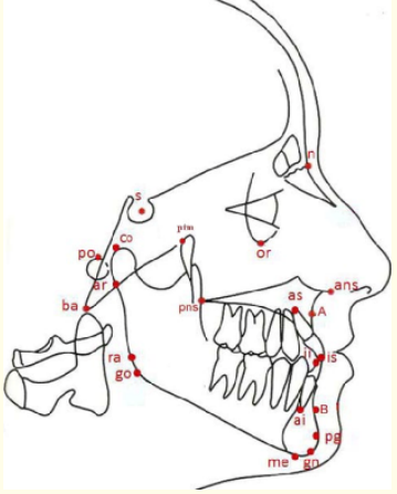
Figure 1: Cephalometric landmarks.
Landmark Abbreviation Definition
Definition of skeletal cephalometric landmarks.
Table
Linear and Angular Measurements
Basal Sagittal measurements
Angular: SNA, SNB, ANB, A:ss-n-Pg, S-N-Pg
Linear: A-NPog
Basal Vertical measurements
Angular: Jaw relationship, Maxillary Inclination, Jaw Angle, Mandibular Inclination,
SN-PoOr.
Linear: PFH, AFH, PFH/AFH, N:ans, Ans:Gn
Mandibular Length
Linear: Go-Co, Co-Gn
Dental measurements
Angular: U1-NL, U1- Inclination, L1-Inclination, U1-L1.
Linear: L1-APog, U1-NA, L1-NB, U1-Protrusion.
Cranial Base Measurements
Angular: n-s-ar, n-s-ba,
Linear: S:Ba, PCB, ACB.
Statistical Analysis
Both Descriptive and Inferential statistics were employed in this study. The level of statistical significance was predetermined at the 0.05 level of confidence. Descriptive statistics that included mean, S.D, Standard Error, 95% confidence interval for mean, Minimum, Maximum values were calculated for each parameter. Inferential Statistics - Independent sample t test between Males and females were compared by using Independent student t test. The mean difference was recorded. The Statistical analysis was done using SPS software version 20.
Studying craniofacial relations and their variations amongst various ethnic groups has long been a subject of investigation. Estab-lishing norms for different racial groups is very helpful in prediction, diagnosis and treatment planning. Many studies have concluded that the standard measurements of one racial group cannot be considered for other racial groups [11,12]. In the Indian scenario, the amount of cephalometric studies to establish norms for Indian people are very scanty and hence have been using western norms which would not be relevant to the Indian population [13-15].
Thus the purpose of this study was to establish lateral cephalometric craniofacial growth changes at 7 years, and also to evaluate possible differences between the genders.
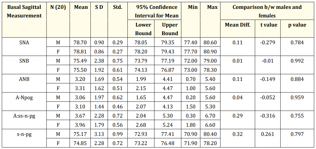
Table 1: Basal Sagittal Measurements at 7 Years of age.
*p < 0.05 and **p < 0.01; Significant ***p < 0.001 Highly Significant; p > 0.05 not significant.
The results of Table 1 inferred that the Mean Sagittal Measurements of 7 year old, when compared between males and females, the females showed a higher values than males for the variables SNA, SNB, ANB, A-Npog, A:ss-n-pg. The mean difference for the variables was very small, hence the p value suggested, that there was no statistically significant difference between the genders for Basal Sagittal Measurements. Hence gender did not have any influence over Basal Sagittal Measurements.
The mean values of SNA for males was 78.70 ± 0.90 and for females was 78.81 ± 0.86, SNB for males was 75.49 ± 2.38 for females was 75.50 ± 1.92. The mean value of ANB for males was 3.20 ± 1.69 and females were 3.31 ± 0.11. These values suggested that though females had higher values for SNA and SNB the value of ANB should have decreased. But ANB also had a higher value suggesting that females had retrognathic mandible than males. This was supported by the mean value of s-n-pg which was 75.17 ± 3.13 for males and 74.85 ± 2.28 for females.
In the previous studies of Chandranee [16] who studied on Chandigarh population of age group of 4-5 years recorded SNA to be of 80.48 ± 2.95 and also in a study by Savadi SC [17] on Karnataka population was found to be 81.70 for the age group of 6-9 years and 84.00 for 10-13 years age. Our study values were in agreement with Savadi SC, but our values were closely matching with Chandranee study. Chandranee [16] who studied on Chandigarh population of age group of 4-5 years recorded mean SNB to be of 75.41 ± 2.77 and also in a study by Savadi SC [17] on Karnataka population was found to be 75.7 for the age group of 6-9 years and 79.3 for 10-13 years age. Our study values were in agreement with Savadi SC, but our values were closely matching with Chandranee study. Savadi SC [17] recorded ANB to be 5.75 for 6-9 years age group and 4.35 for 10-13 years age group.
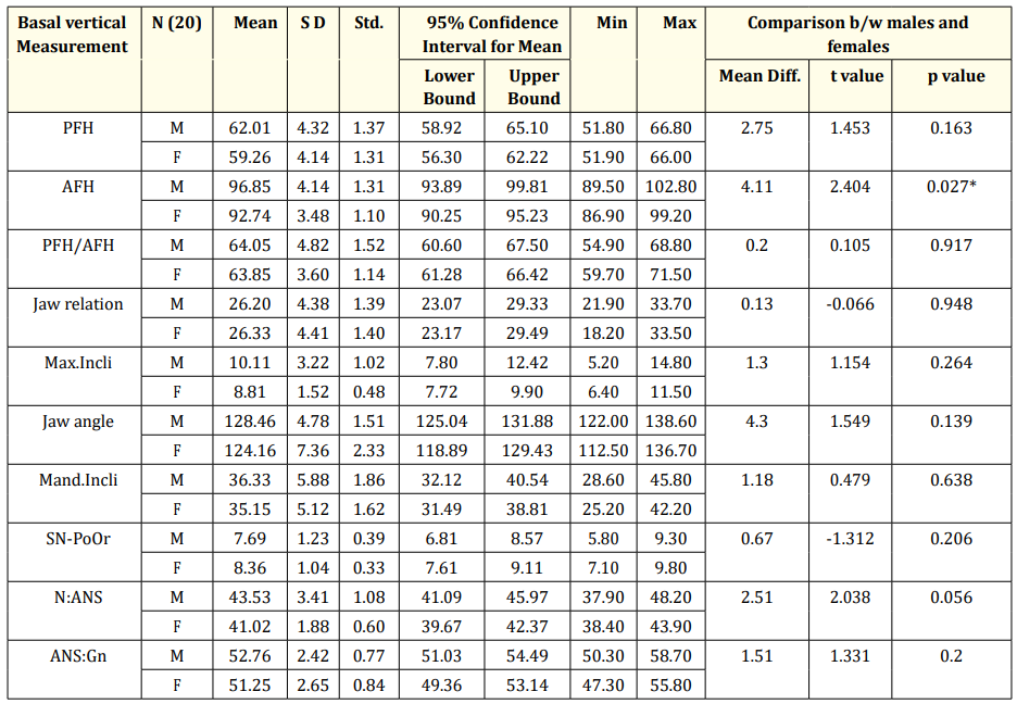
Table 2: Basal Vertical Measurements at 7 Years of age.
*p < 0.05 and **p < 0.01; Significant ***p < 0.001 Highly Significant; p > 0.05 not significant.
The results of Table 2 Inferred that the mean Basal Vertical measurements of 7 year old when compared between Males and fe-males for values of PFH, PFH/AFH, Jaw relation, Maxillary Inclination, Jaw angle, Mandibular Inclination, SN-PoOr & ANS: Gn there was no statistically significant difference between Males and Females.
Whereas the variable AFH, Males of 7 years had a higher value when compared with 7 years old females. The mean AFH at 7 years for Males was 96.85 ± 4.14 and for females was 92.74 ± 3.48 and the mean difference was found to be 4.11 which were statistically sig-nificant. The variable N:ANS Males of 7 years had a higher value when compared with 7 years old females. The mean N:ANS at 7 years for Males was 43.53 ± 3.41 and for females was 41.02 ± 1.88 and the mean difference was found to be 2.51 which was statistically not significant, but the p value was 0.056 close to significance value.
The statistically significant difference in 7 years for the above two variables AFH and N:ANS between Males and females suggest that the boys had relatively Increased Anterior Facial height and upper anterior facial height due to growth of maxillary complex & growth of mandible respectively. AFH and N:ANS which is upper facial height, had a correlation that both were showing increase.
The Mean values of males were higher than the females for PFH, AFH, PFH/AFH, Maxillary Inclination, Jaw Angle, Mandibular Inclination, N:ANS & ANS:Gn, whereas the females showed Higher values for Jaw relation and SN-PoOr.
The mean Mandibular Inclination in a study by Chandranee [16] on age group of 4-5 years was found to be 32.03 ± 3.64 and by Savadi.S.C. [17] On age group of 6-9 years was found to 34.75 and 29.9 at 10-13 years and in our study it was found to be 32.26 ± 4.15 at 7 years.
The results of Table 3 Inferred that mean Mandibular length measurements Go-Co and Co-Gn of 7 year old when compared between males and females, the males had higher mean values than the females for both the parameters. But this higher Mean values were not significant when compared between Males and females.
The above changes in 7 years of variables Go-Co and Co-Gn suggest that Males had increased growth in ramus height and length of Mandible, when compared with females. This suggests that males had a prominent mandible than females at 7 years.
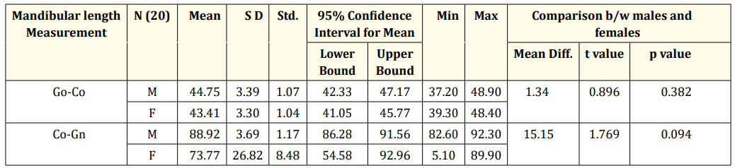
Table 3: Mandibular length Measurements - at 7 Years of age.
*p < 0.05 and **p < 0.01; Significant ***p < 0.001 Highly Significant; p > 0.05 not significant.
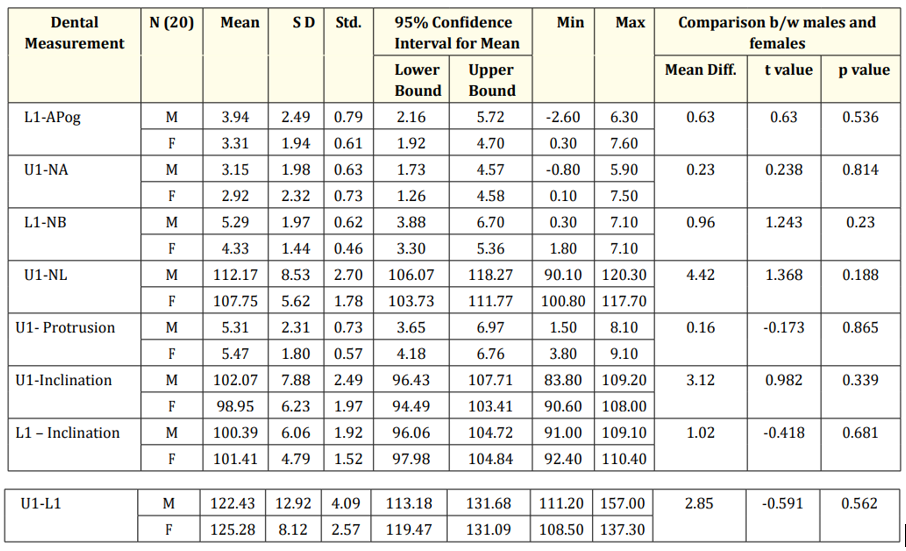
Table 4: Dental Measurements at 7 Years of age.
*p < 0.05 and **p < 0.01; Significant ***p < 0.001 Highly Significant; p > 0.05 not significant.
The results of Table 4 Inferred that the Mean Dental measurements of 7 year old when compared between Males and females for values of L1-APog, U1-NA , L1-NB, U1-NL, U1-Protrusion, U1-Inclination, L1-Inclination and U1-L1, there was differences in the mean values but it was not a statistically significant difference between Males and females for any variables. The Mean values of L1-APog, U1-NA, L1-NB, U1-NL & U1-Inclination showed higher values for males than females, whereas females had higher mean values than males for U1-Protrusion, L1-Inclination & U1-L1.
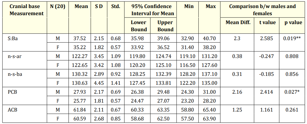
Table 5: Dental Measurements at 7 Years of age.
*p < 0.05 and **p < 0.01; Significant ***p < 0.001 Highly Significant; p > 0.05 not significant.
The mean U1-NA at 7 years was 3.04 ± 2.10 in our study; where as in the studies of Chandranee [16] It was found to be 0.14 ± 1.71 for 4-5 years of age group for Chandigar population. Savadi SC [17] found U1-NA to be 2.15 ± 5.3 for age group of 6-9 years of Karnataka population. The study of Savadi agreed with our study whereas the study of Chandranee was not in agreement because the age of the subjects was very less i.e. 4-5 years. The Mean L1-NB at 7 years was 4.81 ± 1.75 where as in studies of Savadi SC [17] L1-NB was found to be 4.85 ± 6.0 for the age group of 6-9 years of Karnataka population, and study by Chandranee [16] found L1-NB to be 2.80 ± 1.31 for age group of 4-5 years on Chandigarh population. L1 inclination in our study at 7 years was 100.90 ± 5.34, whereas in study by Savadi SC [17] on 6-9 years found to be 93.65 and at 10-13 years to be 100.00 for Karnataka population, this suggested that L1-Inclination was influenced by age and was in agreement with Savadi SC study. U1-L1 in our study, at 7 years was found to be 123.86 ± 10.61, Savadi SC [17] in their study for age group of 6-9 years of Karnataka population found U1-L1 to be 131.9 was not matching with our study. DN Kapoor in a study on Lucknow Population of 6 years was 138.5 ± 9.85 and at 9 years was 125.33 ± 7.49.
The Mean cranial base measurements of 7 year old when compared between Males and females, the males had higher Mean values than the females for S:Ba, PCB & ACB, but females had higher mean values than males for n-s-ar & n-s-ba. The variable S:Ba showed statistically significant difference, where Males had a higher Mean value for S:Ba. The mean S:Ba at 7 years for Males was 37.52 ± 2.15 and for females was 35.22 ± 1.82; the mean difference was 2.3 which was statistically significant. Males had a higher linear measure-ment in S:Ba when compared between genders. The mean PCB at 7 years for Males was 27.93 ± 2.17 and for females was 25.77 ± 1.81; the mean difference was 2.16 which were statistically significant, suggesting that Males had a higher linear measurement in Posterior Cranial Base when compared between genders. OP Kharabanda [18] and Ashima Valiathan [19] in their study suggested that gender did not have any influence on the variable n-s-ar this was matching with our study.
Craniofacial dimensions seen in this study is very useful data of Craniofacial changes of Rural Residential School children of Suttur, but needs to be done on a larger sample.
Copyright: © 2017 Ravi S., et al. This is an open-access article distributed under the terms of the Creative Commons Attribution License, which permits unrestricted use, distribution, and reproduction in any medium, provided the original author and source are credited.


 Open Access by
Acta Scientific is licensed under a Creative Commons Attribution 4.0 International License
Open Access by
Acta Scientific is licensed under a Creative Commons Attribution 4.0 International License
Based on a work at https://actascientific.com
ff
ff
© 2024 Acta Scientific, All rights reserved.