Pedro Reis Conceição*, B Caldeira, J Cortês, M Cotovio, M Rente, I Matias, I Pereira, M Evans, A Laranjeira, J Patrício, R Félix and M Carvalho
Cirurgia Geral, Unidade Local de Saúde do Alentejo Central, Évora, Portugal
*Corresponding Author: Pedro Reis Conceição, Cirurgia Geral, Unidade Local de Saúde do Alentejo Central, Évora, Portugal.
Received: July 10, 2024; Published: September 13, 2024
Citation: Pedro Reis Conceição., et al. “Stump Appendicitis: A Rare but Real Complication". Acta Scientific Clinical Case Reports 8.10 (2024):18-20.
Stump appendicitis can occur as a medium/late complication of a previous appendectomy, and mimics the clinical manifestations of an acute appendicitis, providing a clinical challenge for the clinicians. There are few cases reported in the literature, being a rare condition. We present a clinical case of a man who presented to the emergency department with pain on his right lower quadrant and had been interventioned (appendectomy) 4 months previously. Computed tomography helped diagnosing a stump appendicitis, and he was re-interventioned. Histology confirmed the diagnosis.
The outcome of this condition is generally good when the surgery is performed before major complications.
Keywords: Stump; Appendicitis; Stump Appendicitis; Laparoscopy; Appendectomy; Mid/Late-Term Complication
CT: Computed Tomography; ED: Emergency Department; RLQ: Right Lower Quadrant
Stump appendicitis, an unusual complication of appendectomy [1], is the inflammation of the residual appendix months/years after appendectomy. It occurs in ~1/50,000 cases and it is thought to have increased after the introduction of the laparoscopic technique [2,3] with very few cases described in the literature [4].
The clinical findings are similar to those found in acute appendicitis, and so it poses diagnostic challenges due to its atypical symptoms and presentation.
The diagnosis is made by ultrasound or computed tomography (CT) [3] – in which it is possible to characterize the presence of complications such as abscesses, perforation and peritonitis [5].
The treatment of appendicitis of the appendicular stump involves a new appendectomy, with removal of the remaining appendix.
The prognosis of this pathology depends on the correct diagnosis, with an excellent prognosis when the patient is re-interventioned.
As we can see, stump appendicitis can result in perforation of the stump with intra-abdominal infection, peritonitis, septicemia and eventually death [6]. Therefore, our aim is to shed light in this rare condition and raise awareness of the occurrence of stump appendicitis as a medium/long-term complication of laparoscopic appendectomy.
We present a clinical case of stump appendicitis in a patient who had undergone a previous laparoscopic appendectomy and came to the emergency department (ED) due to pain in the right lower quadrant (RLQ).
A 44-year-old patient came to the ED with abdominal pain located in the RLQ, which had been evolving for 24 hours, with dysuria and fever (38ºC). The patient had a laparoscopic appendectomy 4 months previously.
During laparoscopic surgery, a retrocecal appendicular stump measuring approximately 5 cm was identified.
The base of the appendix was dissected and transsected with EndoGIA Stapler. Histology was compatible with stump appendicitis (Figure 2).
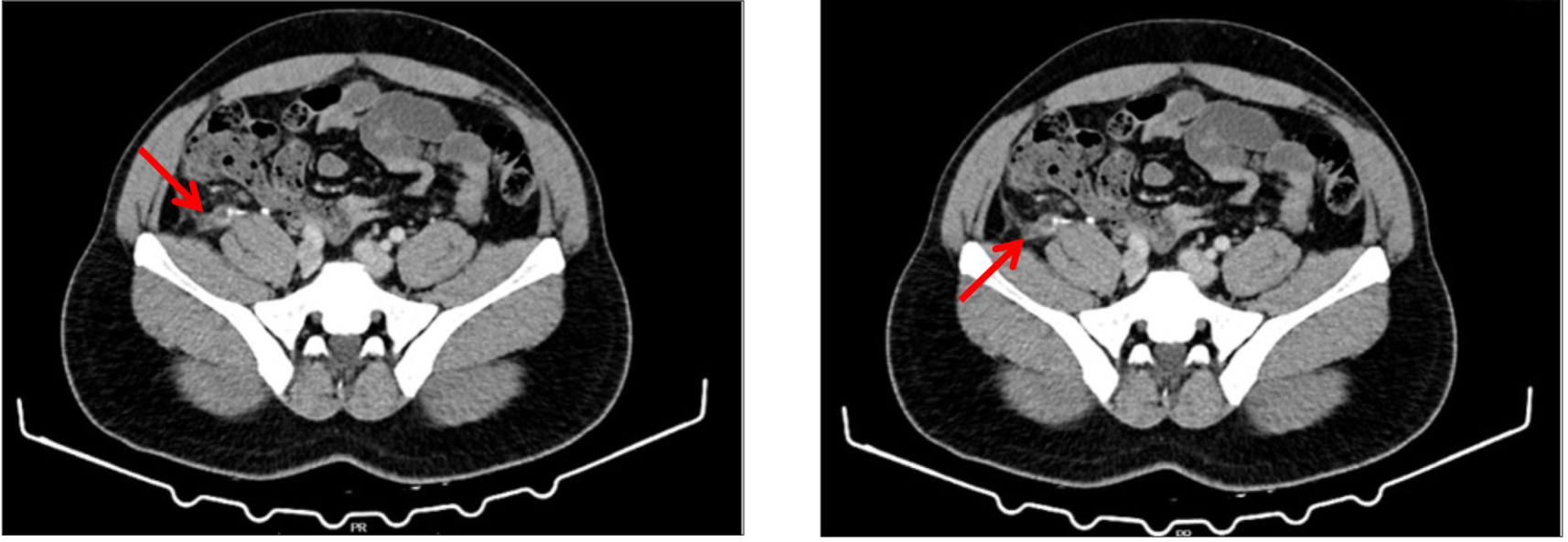
Figure 1: Growth Parameters of Chilli under foliar application of treatments
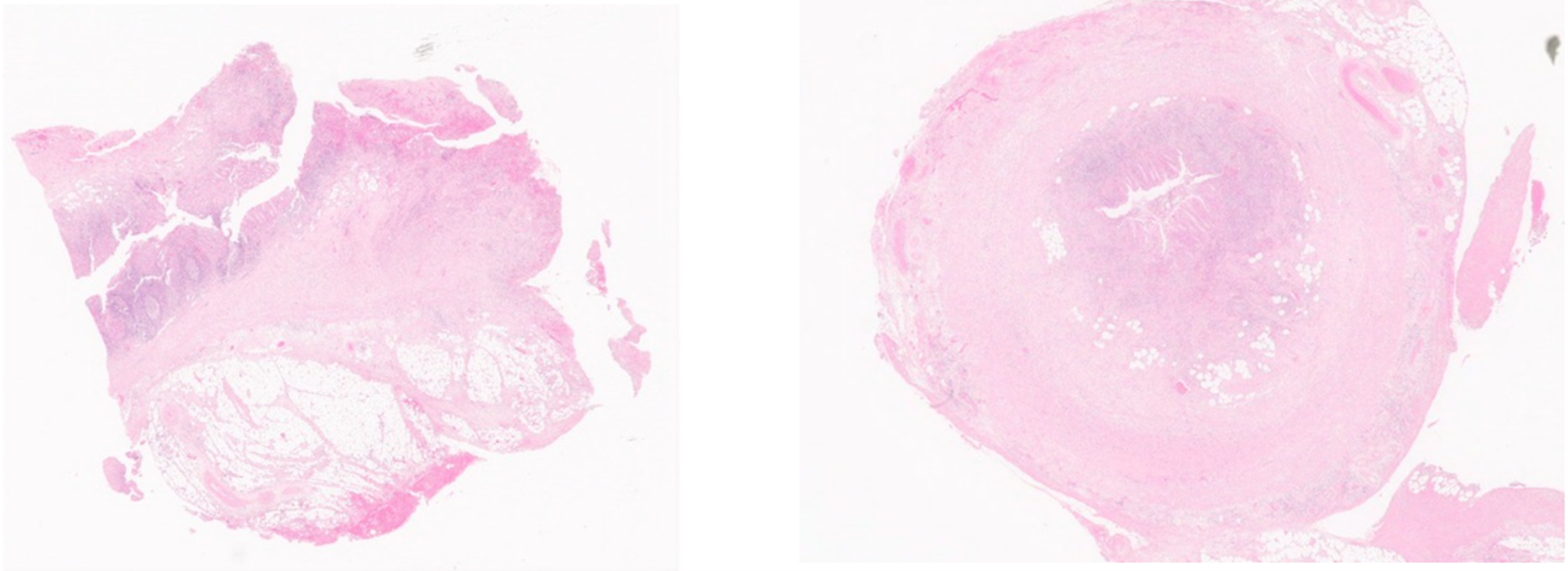
Figure 2: Histology of the specimen. H&E staining. Chronic inflammatory and transmural granulocyte infiltrate with vascular congestion and hemorrhage. Periappendicitis lesions can be seen.
Stump appendicitis is a rare but significant variant of appendicitis. Clinicians should maintain a high index of suspicion in patients presenting with typical abdominal symptoms even after a previous appendectomy. Treatment involves new surgery, with removal of the remaining appendix.
As a prevention, it is suggested to avoid leaving a long appendicular stump when performing an appendectomy – no more than 0.5 cm from its junction to the cecum [4,7]. The outcome is generally good when the surgery is performed before major complications, emphasizing the importance of early diagnosis and intervention.
There is no financial interest or any conflict of interest.
Copyright: © 2024 Pedro Reis Conceição., et al. This is an open-access article distributed under the terms of the Creative Commons Attribution License, which permits unrestricted use, distribution, and reproduction in any medium, provided the original author and source are credited.
 Open Access by
Acta Scientific is licensed under a Creative Commons Attribution 4.0 International License
Open Access by
Acta Scientific is licensed under a Creative Commons Attribution 4.0 International License
Based on a work at https://actascientific.com
ff


